
Head and Neck MRI – Safe and Accurate Diagnosis
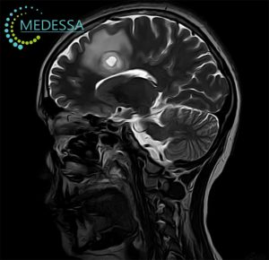
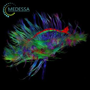
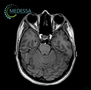
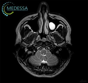
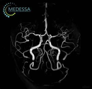
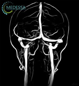
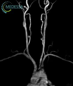
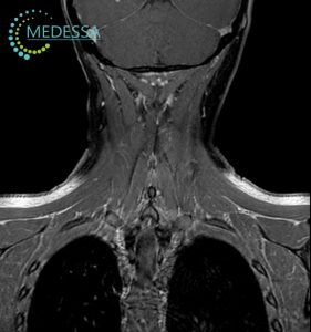
MRI is the most effective method for examining the brain. It is not only safe and non-invasive, but also provides detailed and precise information without exposure to harmful radiation, unlike X-rays or CT scans. Brain MRI allows assessment of all brain structures, including blood vessels, meninges, and nerve roots.
This method enables early detection of various conditions such as tumors, metastases, inflammatory processes, degenerative changes, deformations, and other pathologies, allowing doctors to make a precise diagnosis and prescribe effective treatment.
Advantages of modern MRI for head examination:
Modern MRI machines and specialized protocols maximize the informativeness of the study.
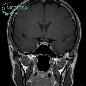
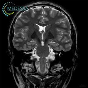
To detect even minor changes, 3D sequences and high-resolution slices are used.
Patient comfort and speed of the procedure are ensured by using a receiver coil integrated into the scanner table, eliminating the need for repositioning.
As a result, doctors receive the most accurate information about early-stage pathologies, including tumors, in a relatively short time. The total scan time for the head without contrast is approximately 10-15 minutes.

Odesa
Phones:
Messengers:
Address: Odesa, 10 Kosmonavtiv Street
Working hours: 8:00 AM – 10:00 PM
Chornomorsk
Phones:
Messengers:
Address: Chornomorsk, 6 Oleksandriyska Street
Working hours: 8:00 AM – 8:00 PM
Primary indications for brain MRI:
- Headaches, acute or chronic;
- Migraines and epileptic seizures;
- Head injuries and consequences of traumatic brain injuries;
- Behavioral changes or psychiatric disorders;
- Dizziness, balance or coordination disturbances;
- Suspicion of tumors or other mass lesions;
- Suspicion of cerebrovascular disorders (strokes, hemorrhages);
- Inflammatory processes, such as meningitis or encephalitis;
- Degenerative brain diseases, such as Parkinson’s or Alzheimer’s;
- Suspicion of demyelinating diseases, such as multiple sclerosis.
When MRI with contrast may be recommended:
- Suspected demyelinating or autoimmune processes to assess inflammation activity;
- Detection of tumors and other mass lesions for better visualization;
- Assessment of blood flow in specific brain regions, including suspected vascular disorders;
- Detection of inflammatory brain conditions such as meningitis or encephalitis;
- Monitoring disease progression, including tumors and degenerative changes;
- Pre-surgical planning to determine the exact location of abnormalities.
Preparation for brain MRI:
- Medical history: Provide information about prior surgeries, chronic illnesses, and medications.
- Removal of metal objects: Remove jewelry, glasses, bracelets, and other metal items.
- Limiting stimulants: Avoid caffeine and other stimulants before the procedure.
- Contrast agent: Blood tests may be required if contrast is planned.
- Consultation with a doctor: Discuss any questions or concerns with your physician before the MRI.
How brain MRI is performed:
During the MRI, the patient lies on a movable table that slides into the scanner. Remaining completely still is essential for obtaining high-quality images. Earplugs or headphones are provided for comfort. A qualified MRI technician monitors the procedure and maintains communication via an internal intercom.
Contraindications for MRI:
MRI is considered safe, but there are absolute and relative contraindications, mostly related to the presence of metal objects or implants.
- Pacemakers or implanted defibrillators;
- Vascular clips or metallic foreign bodies in the brain;
- Implanted neurostimulators or hearing implants;
- Ferromagnetic prostheses or metal fixations;
- Other devices containing metal.
The final decision on performing an MRI is made by the physician.