


MRI 1.5 Tesla
FIRST IN
CHORNOMORSK
The center is equipped with a modern Philips Achieva 1.5 Tesla magnetic resonance imaging system, providing high diagnostic quality.

PROFESSIONALS WITH MANY YEARS OF EXPERIENCE
Our team of highly qualified
like-minded physicians and
caring staff is our pride.
We work together for your
comfort and safety

YOUR COMFORT IS
OUR TOP PRIORITY
Bright and comfortable premises with minimal waiting time.
We do everything to ensure every stage
of the MRI is comfortable for you.

AFFORDABLE PRICING
FOR EVERYONE
MRI 1.5
Affordable prices that
match the high level and
quality of our examinations
MRI in Chornomorsk
Our Doctors

Bondar Eleonora Leonidivna
Radiologist of the Highest Qualification Category
Professional Experience
- Over 35 years of professional experience
- Specialist in MRI and CT diagnostics
- Member of the Ukrainian and European Radiological Associations
- Primary focus — oncology diagnostics and monitoring

Kovalyov Borys Oleksandrovych
Radiologist of the Highest Qualification Category
Professional Experience
- Over 45 years of professional experience
- Specialist in MRI and CT diagnostics
- Member of the Ukrainian and European Radiological Associations
- One of the first MRI and CT diagnostic physicians in Odesa

Borozenko Kseniia Olehivna
Radiologist of the Highest Qualification Category
Professional Experience
- Over 20 years of professional experience
- Specialist in MRI and CT diagnostics
- Member of the Ukrainian and European Radiological Associations
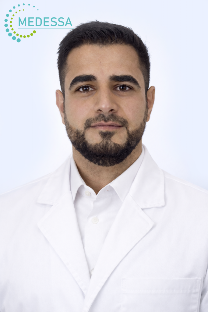
Al-Gburi Ahmed
Radiologist, Clinical Resident specializing in Radiology
Professional Experience
- Over 5 years of professional experience
- Specialist in MRI, CT, and X-ray diagnostics
- Member of the Ukrainian and European Radiological Associations
- Fluent in 4 languages
MRI in Chornomorsk – advanced diagnostic imaging at MEDESSA
About MEDESSA Medical Center
MEDESSA Medical Center in Chornomorsk provides high-quality magnetic resonance imaging (MRI) using a modern 1.5 Tesla scanner. This field strength is considered the international clinical standard, allowing detailed visualization of organs, soft tissues, joints, and the nervous system without ionizing radiation.
MRI 1.5 Tesla systems are widely used in leading hospitals across Europe, the United States, and Asia due to their optimal balance between image quality, diagnostic accuracy, and patient comfort. Examinations are performed according to modern clinical protocols, helping physicians detect diseases early and plan effective treatment strategies.
What is Magnetic Resonance Imaging (MRI)
MRI is a non-invasive diagnostic method that produces highly detailed cross-sectional images of internal anatomical structures. Unlike CT or X-ray imaging, MRI does not use ionizing radiation, making it a safe option even when repeated examinations are required.
MRI is commonly used to evaluate:
- brain and central nervous system
- spine and spinal cord
- joints, ligaments, cartilage, and muscles
- abdominal organs
- pelvic organs
- soft tissues
- vascular structures
- endocrine organs
MRI can detect inflammatory conditions, tumors, degenerative diseases, traumatic injuries, vascular abnormalities, congenital conditions, and many other clinically significant findings.
How MRI works
MRI technology uses a strong magnetic field combined with radiofrequency pulses to generate high-resolution images of internal tissues. The scanner produces detailed cross-sectional images that allow physicians to assess anatomical structures with high precision.
Due to excellent soft tissue contrast, MRI is considered one of the most informative imaging techniques in neurology, orthopedics, oncology, gastroenterology, urology, and many other medical specialties.
Benefits of 1.5 Tesla MRI in Chornomorsk
The 1.5 Tesla MRI scanner represents a globally accepted diagnostic standard, providing high-quality imaging for the majority of clinical indications. It allows accurate detection of structural abnormalities and supports early diagnosis.
- high-resolution imaging of anatomical structures
- early detection of pathological changes
- non-invasive and painless procedure
- no exposure to ionizing radiation
- internationally accepted clinical protocols
- comfortable examination conditions
1.5 Tesla MRI is highly effective for imaging the brain, spine, joints, abdominal organs, pelvic organs, vascular system, and soft tissues.
Why choose MEDESSA
MEDESSA in Chornomorsk combines modern technology with experienced medical specialists and evidence-based imaging protocols. Accurate MRI results help physicians establish diagnoses, monitor disease progression, and select the most appropriate treatment approach.
Timely MRI examination helps:
- detect diseases at early stages
- confirm or уточнить diagnosis
- monitor treatment effectiveness
- prepare for surgical procedures
- reduce risk of complications
Schedule your MRI in Chornomorsk at MEDESSA and receive accurate diagnostic results aligned with modern international medical standards.
1.5 Tesla MRI in Chornomorsk – reliable diagnostic standard
About 1.5 Tesla MRI
MEDESSA Medical Center in Chornomorsk provides magnetic resonance imaging using a modern 1.5 Tesla MRI scanner. This field strength is considered the global clinical standard and is widely used in hospitals across Europe, the United States, and Asia due to its optimal balance of diagnostic accuracy and patient comfort.
1.5 Tesla MRI produces detailed cross-sectional images of internal organs, soft tissues, joints, and the nervous system. The method allows physicians to evaluate anatomical structures, detect pathological changes, and determine the most appropriate treatment strategy. MRI does not use ionizing radiation and is considered a safe diagnostic technique.
Benefits of 1.5 Tesla MRI
- 1. High image quality: provides detailed visualization of soft tissues, organs, joints, vascular structures, and nervous system components.
- 2. Accurate disease detection: MRI helps identify tumors, inflammatory processes, degenerative disorders, traumatic injuries, vascular abnormalities, and congenital conditions.
- 3. Advanced brain imaging: enables assessment of cerebral cortex, white matter, pituitary gland, cerebellum, brainstem, cerebrospinal fluid spaces, and vascular structures.
- 4. Effective spine and joint evaluation: intervertebral discs, cartilage, ligaments, tendons, muscles, and nerve roots are clearly visualized.
- 5. Reliable imaging of internal organs: MRI is used to evaluate liver, kidneys, pancreas, pelvic organs, lymph nodes, and soft tissues.
- 6. Safe and non-invasive procedure: MRI does not involve radiation exposure and can be repeated when clinically necessary.
- 7. Advanced imaging protocols: MR angiography and specialized imaging sequences allow comprehensive assessment of anatomical regions.
Why 1.5 Tesla MRI is considered the clinical standard
1.5 Tesla MRI provides highly accurate visualization for most clinical indications and is widely recommended in international medical guidelines. The method allows early detection of pathological changes and supports monitoring of treatment outcomes.
MRI is extensively used in neurology, orthopedics, oncology, traumatology, urology, gynecology, and gastroenterology due to its excellent soft tissue contrast and diagnostic value.
MRI in Chornomorsk at MEDESSA
At MEDESSA in Chornomorsk, MRI examinations are performed according to modern international imaging protocols. Optimized scanning parameters allow accurate and clinically useful diagnostic results.
Timely MRI examination helps detect diseases at early stages, confirm diagnosis, monitor treatment effectiveness, and reduce the risk of complications. Schedule your MRI examination at MEDESSA to receive high-quality diagnostic imaging.
Contrast-Enhanced MRI in Chornomorsk 1.5 Tesla
Contrast enhancement in 1.5 Tesla MRI
Contrast-enhanced magnetic resonance imaging is an important additional step used in selected clinical situations to improve diagnostic accuracy. At MEDESSA Medical Center in Chornomorsk, contrast-enhanced MRI is performed on a modern 1.5 Tesla scanner, which is considered the international clinical standard for MRI and provides highly informative imaging of the brain, spine, joints, soft tissues, internal organs, and vascular structures.
During the examination, a gadolinium-based contrast agent may be administered intravenously when clinically indicated. Contrast enhancement helps differentiate normal tissue from abnormal tissue more clearly, evaluate vascularity, improve lesion characterization, detect inflammatory activity, assess blood-brain barrier disruption, identify postoperative changes, and refine the evaluation of many complex pathological processes.
When contrast-enhanced MRI is especially useful
Contrast is not required for every MRI examination. However, in many clinical scenarios it significantly increases the diagnostic value of the study. The decision to use contrast is made individually based on symptoms, suspected diagnosis, anatomical region, and the specific clinical question.
- Tumors and mass lesions: contrast helps define lesion borders, internal structure, enhancement pattern, and relationship to surrounding tissues.
- Inflammatory and infectious disorders: it improves visualization of active inflammatory changes in the brain, spine, soft tissues, and internal organs.
- Vascular abnormalities: contrast may improve assessment of perfusion, selected vascular malformations, and consequences of impaired circulation.
- Demyelinating diseases: it is valuable for assessing lesion activity in conditions such as multiple sclerosis and other central nervous system disorders.
- Postoperative follow-up: contrast-enhanced MRI may help distinguish postoperative scar tissue from residual or recurrent disease.
- Complex diagnostic cases: it is particularly helpful when non-contrast MRI alone does not provide enough clinically relevant information.
Benefits of contrast-enhanced 1.5 Tesla MRI
- 1. Higher diagnostic accuracy: contrast enhancement improves lesion detection and characterization in many clinical situations.
- 2. Better tissue and vascular visualization: it provides additional information about lesion boundaries, blood supply, and biological activity.
- 3. Improved differential diagnosis: contrast-enhanced MRI helps distinguish inflammatory, neoplastic, vascular, and postoperative changes more precisely.
- 4. Support for treatment planning: findings may guide therapy selection, follow-up strategy, surgical planning, and monitoring of treatment response.
- 5. Broad clinical usefulness: the method is widely applied in neurology, oncology, orthopedics, urology, gynecology, gastroenterology, and other medical specialties.
Safety of gadolinium-based contrast agents
Modern gadolinium-based contrast agents are generally well tolerated, and serious adverse reactions are uncommon. Even so, contrast administration should always be considered individually, with attention to kidney function, allergy history, previous reactions to contrast agents, pregnancy status, and other relevant clinical factors.
Before the examination, the medical team may ask about:
- kidney disease or reduced kidney function
- previous allergic reactions
- prior reactions to contrast agents
- pregnancy or other clinically important conditions
In patients with significantly impaired renal function, the decision to administer contrast requires careful risk assessment. For this reason, contrast-enhanced MRI should always be used according to medical indications and individualized safety considerations.
Why choose MEDESSA in Chornomorsk
At MEDESSA in Chornomorsk, contrast-enhanced MRI is performed on a 1.5 Tesla scanner using modern imaging protocols designed to deliver detailed and clinically meaningful results. This approach helps physicians not only visualize anatomy, but also better understand the extent, activity, and nature of many disease processes.
Contrast-enhanced MRI is especially valuable when the goal is to achieve a more precise diagnosis and support evidence-based treatment decisions. Accurate imaging plays an important role in early detection, follow-up, and individualized patient care.
Preparation for 1.5 Tesla MRI in Chornomorsk
How to prepare for an MRI examination
Proper preparation for magnetic resonance imaging at MEDESSA Medical Center in Chornomorsk helps ensure clear, high-quality, and clinically useful images. A 1.5 Tesla MRI scanner is considered the international standard in modern MRI diagnostics and is widely used for imaging the brain, spine, joints, soft tissues, blood vessels, abdominal organs, pelvic organs, and retroperitoneal structures.
In many cases, MRI does not require complicated preparation. However, some types of examinations do involve specific recommendations that can improve image quality and make the procedure more effective.
What to do before arriving at the center
It is advisable to arrive in advance so there is enough time for registration, document review, safety screening, and clarification of examination details. Before the scan, patients should inform the medical team about implants, pacemakers, vascular clips, metallic hardware, cochlear implants, insulin pumps, neurostimulators, pregnancy, kidney disease, allergy history, or any previous MRI examinations with contrast administration.
Preparation immediately before the procedure
Before entering the MRI room, all metal objects and electronic devices must be removed. This includes jewelry, watches, glasses, hearing aids, bank cards, mobile phones, keys, hairpins, belts, and other accessories. Clothing should be comfortable and should not contain metallic zippers, buttons, underwires, or decorative metal elements.
If the examination involves joints, soft tissues, or a specific anatomical area, it is usually advisable to remove tapes, adhesive patches, braces, elastic supports, bandages, and other external fixation devices unless the physician has provided different instructions. It is also preferable not to apply creams, gels, ointments, or warming agents to the area being examined immediately before the scan.
Which documents are useful to bring
For more accurate interpretation, it is helpful to bring previous medical records when available:
- prior MRI, CT, ultrasound, or X-ray results
- hospital discharge summaries or specialist reports
- the referral for MRI
- information about previous surgeries
- laboratory test results if contrast-enhanced MRI is planned
Comparison with prior studies often helps the radiologist assess disease progression, treatment response, and the nature of structural changes over time.
Why it is important to remain still during MRI
Remaining still during MRI is essential because even minor movement can reduce image quality and limit diagnostic accuracy. The more stable the body position during scanning, the more precise the final images will be.
If a patient has significant pain, difficulty lying still, increased anxiety, or claustrophobia, it is best to mention this in advance. In selected cases, the physician may recommend additional preparation or discuss the possibility of supportive medication before the examination.
Preparation for MRI of the head and brain
Before MRI of the head or brain, it is preferable not to use decorative cosmetics, especially products containing metallic particles or glitter. False eyelashes, metallic hair accessories, and decorative hair elements should also be avoided because they may affect image quality or cause discomfort during the scan.
These recommendations are particularly important for examinations of the brain, orbits, pituitary gland, paranasal sinuses, and other head structures where high-resolution imaging is required.
Preparation for MRI of joints and soft tissues
MRI of joints, muscles, ligaments, tendons, and soft tissues usually does not require special dietary preparation. However, it is important to remove braces, supports, bandages, and other external fixation devices when appropriate, and to avoid applying topical medications to the examination area unless otherwise advised by a physician.
These examinations provide detailed visualization of cartilage, joint surfaces, menisci, ligaments, tendons, muscles, fascia, and periarticular soft tissues, helping detect both common and less typical pathological changes.
Eating and drinking before MRI of the abdomen, pelvis, and retroperitoneal space
Some MRI examinations of the abdominal organs, pelvic organs, or retroperitoneal space may require temporary dietary restrictions. These recommendations are often intended to reduce bowel motion and gas, which can improve image quality.
Depending on the type of study, patients may be advised to:
- avoid food for several hours before the examination
- temporarily limit foods that increase gas formation
- follow specific instructions regarding bladder filling for pelvic MRI
Because preparation can vary depending on the anatomical region and the clinical question, the exact instructions for abdominal MRI, pelvic MRI, retroperitoneal MRI, or contrast-enhanced MRI should be confirmed in advance.
Preparation for contrast-enhanced MRI
If contrast-enhanced MRI is planned, the patient may be asked additional questions about kidney function, allergy history, previous reactions to contrast agents, and associated medical conditions. In some cases, laboratory assessment of kidney function may be recommended before the examination.
Contrast administration is performed only when clinically indicated and may improve diagnostic accuracy in the evaluation of tumors, inflammatory disease, vascular abnormalities, active demyelinating lesions, and other important pathological processes.
MRI in Chornomorsk at MEDESSA
At MEDESSA Medical Center in Chornomorsk, 1.5 Tesla MRI examinations are performed according to modern diagnostic protocols. Proper preparation helps achieve the most accurate results, which are important for diagnosis, treatment planning, and follow-up assessment.