
Spine MRI in Chornomorsk (1.5 Tesla)
Magnetic Resonance Imaging (MRI) of the spine is a highly informative and safe diagnostic method used to evaluate the vertebral column, spinal cord, intervertebral discs, and surrounding soft tissues. MRI does not use ionizing radiation, making it safer than X-ray or CT examinations. This technology is considered the clinical standard for diagnosing degenerative, inflammatory, traumatic, and oncological spinal conditions.
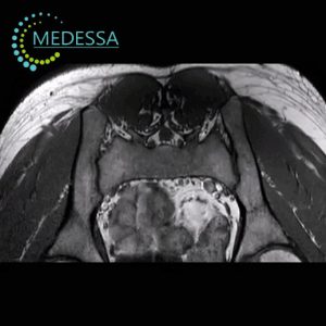
At the MEDESSA medical center in Chornomorsk, spine MRI is performed using a 1.5 Tesla scanner. This field strength is widely recognized as the international diagnostic standard, providing high-quality images of discs, nerves, muscles, ligaments, and spinal cord structures.
Chornomorsk
Phones:
Messengers:
Address: Chornomorsk, 6 Oleksandriyska Street
Working hours: 8:00 AM – 8:00 PM
What Spine MRI Detects
- degenerative disc disease and osteochondrosis;
- disc protrusions and herniations;
- spinal canal stenosis;
- inflammatory diseases of the spine and spinal cord;
- benign and malignant tumors;
- metastatic lesions;
- compression fractures;
- congenital abnormalities;
- multiple sclerosis;
- facet joint arthropathy;
- spinal deformities such as scoliosis or kyphosis.
Anatomical Structures Visualized
- vertebral bodies and bone marrow;
- intervertebral discs;
- spinal cord and nerve roots;
- ligaments and joints;
- paraspinal muscles;
- soft tissues;
- vascular structures.
Indications for Spine MRI
- acute or chronic back pain;
- radiating pain to arms or legs;
- numbness or tingling sensations;
- limited mobility;
- suspected disc herniation;
- spinal trauma;
- suspected tumors;
- treatment monitoring;
- neurological symptoms;
- pelvic organ dysfunction.
MRI of Different Spine Regions
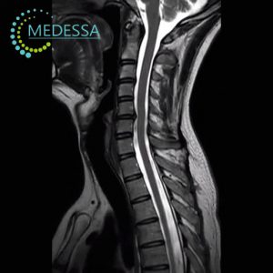
Cervical Spine MRI
- neck pain and stiffness;
- headaches and dizziness;
- arm numbness;
- tinnitus;
- restricted mobility;
- disc pathology;
- vascular compromise affecting the brain.
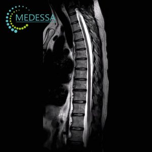
Thoracic Spine MRI
- pain between shoulder blades;
- intercostal neuralgia;
- muscle spasms;
- postural abnormalities;
- pain during breathing.
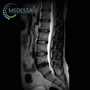
Lumbar Spine MRI
- lower back pain;
- sciatica;
- leg numbness;
- disc degeneration;
- nerve compression.
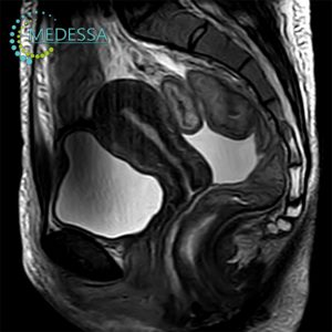
Sacral and Coccygeal MRI
- sacral pain;
- post-traumatic changes;
- inflammatory conditions;
- tumor suspicion;
- neurological symptoms.
Contrast-enhanced MRI
Contrast agents based on gadolinium may be used when additional diagnostic accuracy is required. Contrast improves visualization of tumors, inflammation, vascular abnormalities, and postoperative changes.
Benefits of 1.5 Tesla MRI in Chornomorsk
1.5 Tesla MRI provides excellent diagnostic accuracy and is considered the global standard for spine imaging. It ensures detailed visualization of soft tissues and nerve structures while maintaining patient comfort.
Preparation
- complete a safety questionnaire;
- remove metallic objects;
- inform the doctor about implants;
- fasting may be required for contrast studies.
Procedure
The patient lies on a movable table that slides into the scanner. Remaining still during the scan ensures high image quality. Ear protection is provided for comfort.
Results
Images are provided in DICOM format. A radiologist’s report is typically available within 24 hours.
Contraindications
MRI may be противопоказана in patients with pacemakers, ferromagnetic implants, or certain electronic medical devices.