
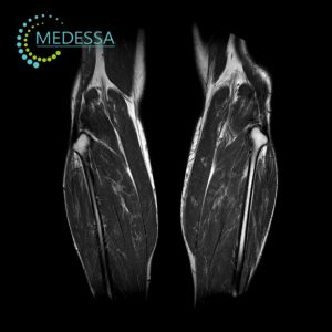
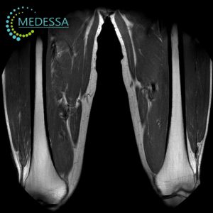
MRI of Joints and Soft Tissues in Chornomorsk (1.5 Tesla)
MRI of joints and soft tissues is a highly accurate diagnostic method used to evaluate the knee, shoulder, hip, ankle, elbow, wrist, and surrounding soft tissue structures. MRI is considered the clinical gold standard for imaging ligaments, tendons, cartilage, menisci, muscles, and synovial membranes.
At MEDESSA in Chornomorsk, examinations are performed using a 1.5 Tesla MRI scanner, providing high-resolution images and accurate assessment of joint pathology.
Chornomorsk
Phones:
Messengers:
Address: Chornomorsk, 6 Oleksandriyska Street
Working hours: 8:00 AM – 8:00 PM
Structures visualized
- articular cartilage;
- menisci;
- ligaments;
- tendons;
- muscles;
- joint capsule;
- synovial membrane;
- bone marrow;
- bursae;
- nerves;
- soft tissues.
What MRI detects
- ligament tears;
- meniscal injuries;
- osteoarthritis;
- arthritis;
- bursitis;
- muscle inflammation;
- tumors;
- cysts;
- traumatic injuries;
- hematomas;
- nerve compression.
Common MRI exams
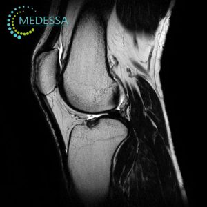
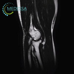
Knee MRI
Evaluates menisci, ligaments, cartilage and muscles.
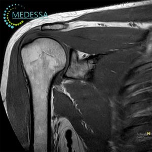
Shoulder MRI
Visualizes rotator cuff tendons and labrum.
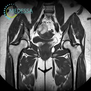
Hip MRI
Assesses cartilage and surrounding soft tissues.
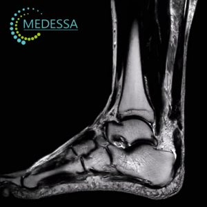
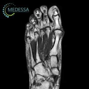
Ankle MRI
Detects ligament tears and tendon injuries.
Soft tissue MRI
Evaluates muscles, fascia, nerves and vessels.
Indications
- joint pain;
- limited mobility;
- swelling;
- trauma;
- suspected ligament tear;
- degenerative disease;
- treatment monitoring;
- suspected tumor.
Contrast MRI
Contrast agents improve visualization of tumors and inflammatory processes.
Benefits of 1.5 Tesla MRI
1.5 Tesla MRI provides high diagnostic accuracy and detailed imaging of joint structures.
Preparation
- remove metal objects;
- inform the doctor about implants;
- fasting may be required for contrast MRI.
Contraindications
- pacemakers;
- metal implants;
- neurostimulators;
- clips;
- electronic devices.