
MRI of the Head and Neck in Chornomorsk — Accurate 1.5 Tesla MRI Diagnostics
MRI of the head and neck is a modern non-invasive imaging method that provides a detailed assessment of the brain, soft tissues of the head and neck, vascular structures, and adjacent anatomical regions. It helps detect a wide range of pathological changes, including early-stage abnormalities. At MEDESSA in Chornomorsk, examinations are performed on a 1.5 Tesla MRI scanner. This field strength is the established clinical standard for routine MRI and offers a reliable balance of image quality, diagnostic value, patient comfort, and examination time for most neurological and soft tissue indications.
Unlike X-ray and CT, MRI does not use ionizing radiation. That is why it is widely used for the diagnosis of neurological, vascular, inflammatory, neoplastic, and degenerative conditions. MRI of the head and neck allows the physician to identify both pronounced structural abnormalities and subtle lesions that may be crucial for making an accurate diagnosis and planning further treatment.

Chornomorsk
Phones:
Messengers:
Address: Chornomorsk, 6 Oleksandriyska Street
Working hours: 8:00 AM – 8:00 PM
What MRI of the Head and Neck Can Show
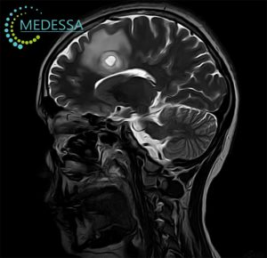
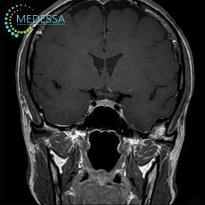
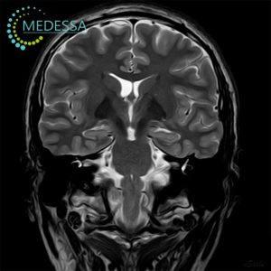
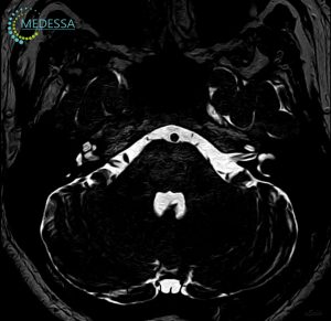
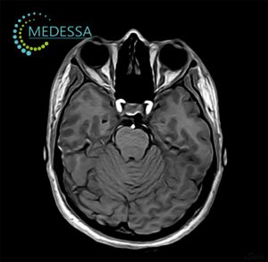
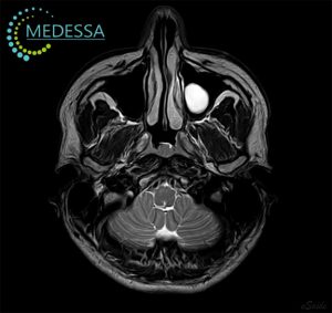
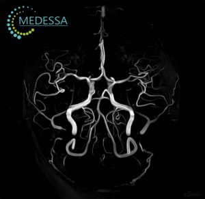
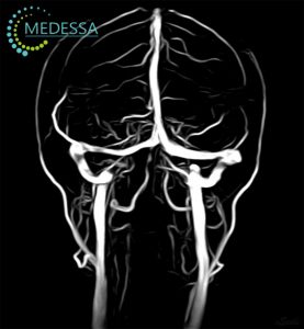
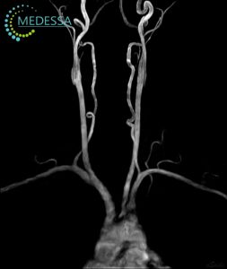
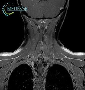
Depending on the clinical indication and scanning protocol, MRI can visualize the cerebral hemispheres, cortex and white matter, ventricular system, corpus callosum, brainstem, cerebellum, pituitary region when indicated, meninges, paranasal sinuses, orbits within the scan area, soft tissues of the neck, salivary glands, lymph nodes, vascular structures, and other relevant anatomical regions.
This examination can help detect ischemic and post-ischemic changes, consequences of traumatic brain injury, intracranial hemorrhage, demyelinating lesions, benign and malignant tumors, metastatic disease, edema, signs of increased intracranial pressure, inflammatory disorders, meningeal involvement, neck masses, and soft tissue pathology associated with both acute and chronic disease.
Why 1.5 Tesla MRI Is a Strong Choice for Most Head and Neck Examinations
1.5 Tesla MRI is a widely accepted standard in modern clinical practice and is used worldwide for imaging the brain, vascular structures, paranasal sinuses, orbits, and soft tissues of the neck. It provides dependable diagnostic performance for most routine and clinically important indications.
For the majority of primary and follow-up neurological examinations, 1.5 Tesla MRI delivers detailed images that are sufficient for identifying both common and less common pathology. For this reason, MRI in Chornomorsk on a 1.5 Tesla scanner is a medically justified and up-to-date solution for high-quality diagnostic assessment.
Main Advantages of MRI of the Head and Neck
- no radiation exposure;
- excellent soft tissue contrast;
- early detection of pathological changes;
- detailed evaluation of the brain and neck soft tissues;
- contrast-enhanced imaging when clinically indicated;
- high diagnostic value in neurological, vascular, oncologic, and inflammatory conditions;
- useful for treatment monitoring and follow-up imaging.
When MRI of the Brain and Neck May Be Recommended
- frequent or persistent headaches;
- migraine, including migraine with aura;
- dizziness, imbalance, or coordination problems;
- episodes of fainting or unexplained loss of consciousness;
- seizures or suspected epilepsy-related conditions;
- memory decline, reduced concentration, or cognitive changes;
- speech problems, limb weakness, or numbness;
- suspected stroke or consequences of impaired cerebral circulation;
- head trauma and post-traumatic follow-up;
- suspected tumor, cyst, or other space-occupying lesion;
- suspected multiple sclerosis or other demyelinating disorders;
- possible meningitis, encephalitis, or other inflammatory disease;
- painful or enlarged structures in the neck;
- enlarged lymph nodes;
- clarification of abnormalities seen on other imaging studies.
Which Conditions MRI Can Help Detect
MRI of the head and neck is used to evaluate a broad range of disorders. Common findings include ischemic lesions, post-traumatic changes, chronic microvascular abnormalities, cysts, benign and malignant tumors, metastatic involvement, inflammatory disease, complicated sinus pathology, soft tissue lesions of the neck, congenital abnormalities, sequelae of neuroinfection, and demyelinating changes associated with suspected multiple sclerosis.
In more complex cases, MRI helps define the exact location of the pathological process, its extent, its relationship to adjacent anatomical structures, and changes over time during treatment. This is especially important for neurologists, neurosurgeons, oncologists, ENT specialists, and other physicians involved in patient management.
When Contrast-Enhanced MRI May Be Needed
Contrast is not required for every MRI examination. Gadolinium-based contrast agents are used only when there is a clear clinical indication. Contrast-enhanced MRI may be recommended for more accurate evaluation of tumors, inflammatory activity, blood-brain barrier disruption, postoperative changes, and selected vascular or demyelinating lesions.
- suspected primary or secondary brain tumors;
- further characterization of a mass lesion;
- assessment of lesion activity in demyelinating disease;
- evaluation of inflammatory disorders of the central nervous system;
- postoperative follow-up imaging;
- differentiation between scar tissue and active disease;
- more precise assessment of pathology involving the head and neck region.
The need for contrast is determined individually by the physician. In patients with kidney disease or other risk factors, additional evaluation may be required before contrast administration.
Preparation for MRI of the Head and Neck
- Medical history: before the scan, it is important to inform the staff about previous surgeries, implanted devices, pacemakers, vascular clips, hearing implants, neurostimulators, metal fragments, chronic illnesses, and prior imaging results.
- Documents: it is advisable to bring your referral, medical reports, and previous MRI, CT, ultrasound, or other imaging results if available.
- Metal objects: before entering the MRI room, jewelry, watches, glasses, hearing aids, removable dental appliances, bank cards, and other metal-containing or electronic items must be removed.
- Food and drink: for standard MRI without contrast, no special preparation is usually needed. If contrast is planned, instructions regarding food intake and any necessary laboratory tests are provided individually.
- Comfort considerations: if you have severe anxiety, claustrophobia, or difficulty remaining still, this should be discussed in advance.
How the Procedure Is Performed
During the examination, the patient lies on a movable table that slides into the scanner. Remaining still is essential for obtaining clear diagnostic images. The scanner produces characteristic sounds, so ear protection such as earplugs or headphones is usually provided. Throughout the procedure, the patient remains under staff supervision and can communicate with the MRI technologist through an internal communication system.
The duration of MRI of the head and neck depends on the scope of the study, the clinical question, and whether contrast enhancement is required. A standard examination usually takes about 15 to 30 minutes, while extended protocols may take longer.
Contraindications and Limitations
MRI is considered a safe imaging technique, but absolute and relative contraindications must always be assessed before scanning. What matters is not only whether an implant is present, but also its MRI compatibility, device model, material, safety conditions, and the anatomical area being examined.
- MRI-incompatible pacemakers or implanted defibrillators;
- certain neurostimulators, cochlear implants, and electronic devices;
- some ferromagnetic clips, implants, or metallic foreign bodies;
- metal fragments, especially in or near the eyes or vital structures;
- serious patient condition when stillness cannot be maintained;
- selected cases during pregnancy, assessed individually;
- severe claustrophobia without prior planning of examination conditions.
The final decision on whether MRI can be performed is made after a safety interview and an individual risk assessment.
MRI of the Head and Neck in Chornomorsk at MEDESSA
If you need informative MRI of the head and neck in Chornomorsk, MEDESSA provides examinations on a 1.5 Tesla scanner using modern MRI protocols. This helps detect pathology in a timely manner, clarify the diagnosis, support treatment planning, and monitor disease over time.