
MR Angiography of the Brain Arteries in Chornomorsk – Cerebral MRA on Philips Achieva 1.5 Tesla
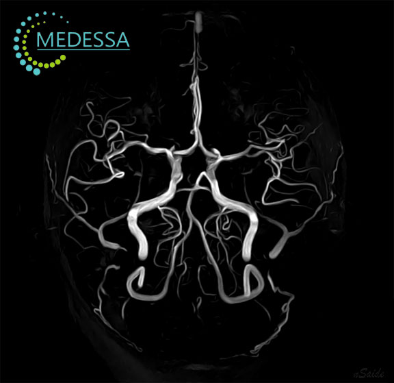
Magnetic resonance angiography of the brain arteries, also known as cerebral MRA, intracranial MRA, or MR angiography of the Circle of Willis, is a specialized neurovascular MRI examination used to visualize the intracranial arterial circulation. It allows assessment of the intracranial internal carotid arteries, vertebral arteries, basilar artery, anterior, middle, and posterior cerebral arteries, communicating arteries, and their major visible branches.
At MEDESSA diagnostic center in Chornomorsk, MR angiography of the brain arteries is performed on a Philips Achieva 1.5 Tesla MRI system. Optimized three-dimensional Time-of-Flight angiography can provide clinically valuable visualization of the cerebral arteries without ionizing radiation and, in many cases, without intravenous contrast administration. The examination may help identify intracranial aneurysms, arterial narrowing, major vascular occlusion, congenital arterial variants, arteriovenous malformations, and other abnormalities of the cerebral circulation.
Modern 1.5 Tesla MRA is an established neurovascular imaging method used worldwide. Its diagnostic effectiveness depends on appropriate protocol selection, arterial flow characteristics, spatial resolution, patient motion control, analysis of original source images, reconstruction quality, and specialist interpretation. When findings remain uncertain or urgent treatment decisions are required, additional CT angiography, digital subtraction angiography, vessel wall MRI, or other vascular imaging may be necessary.
What Is MR Angiography of the Brain Arteries?
MR angiography is a group of magnetic resonance techniques designed to visualize blood vessels and evaluate flow-related vascular anatomy. Cerebral MRA primarily examines the arterial lumen, vascular course, branching pattern, symmetry, caliber, continuity, and flow-related signal within the intracranial arteries.
Depending on the clinical indication, brain artery MRA may be performed:
- without contrast using three-dimensional Time-of-Flight technology;
- with a gadolinium-based contrast agent in selected cases;
- as part of a comprehensive brain MRI protocol;
- together with MR angiography of the neck vessels;
- together with diffusion MRI in suspected stroke or transient ischemic attack;
- for follow-up after selected neurosurgical or endovascular procedures.
MRA is non-invasive and does not require insertion of an arterial catheter. However, it does not completely replace digital subtraction angiography when very high spatial or temporal resolution is required, vascular anatomy remains unclear, detailed hemodynamic evaluation is needed, or an endovascular procedure is being planned.
When Is Cerebral Artery MRA Recommended?
MR angiography of the intracranial arteries may be recommended when symptoms, clinical findings, previous imaging, family history, or known vascular disease suggest abnormal cerebral circulation.
- Suspected intracranial aneurysm;
- Screening in selected patients with an increased hereditary risk of intracranial aneurysm;
- Sudden or unusual severe headache after urgent causes have been appropriately assessed;
- Transient ischemic attack;
- Suspected ischemic stroke;
- Focal neurological deficit;
- Speech disturbance;
- Unilateral weakness or numbness;
- Suspected intracranial arterial stenosis;
- Suspected arterial occlusion;
- Arteriovenous malformation or vascular fistula;
- Suspected cerebral vasculitis or another intracranial arteriopathy;
- Possible arterial dissection;
- Pulsatile tinnitus in selected clinical situations;
- Congenital cerebrovascular abnormality;
- Preoperative neurovascular assessment;
- Follow-up of a known untreated aneurysm;
- Follow-up after aneurysm treatment;
- Monitoring after vascular surgery, stenting, coiling, or another endovascular procedure;
- Evaluation of anatomical variants of the Circle of Willis.
Sudden severe headache, acute weakness, speech disturbance, facial asymmetry, loss of consciousness, or other acute neurological symptoms require urgent emergency evaluation. A scheduled outpatient MRA should not delay emergency stroke or hemorrhage assessment.
How MR Angiography Differs From Standard Brain MRI
MR angiography and conventional brain MRI examine different aspects of the nervous system. MRA primarily evaluates the intracranial arteries and flow-related vascular anatomy. Standard brain MRI evaluates the brain tissue and surrounding intracranial structures.
| Standard Brain MRI | MR Angiography of Brain Arteries |
|---|---|
| Evaluates brain tissue | Evaluates intracranial arteries |
| Shows gray and white matter | Shows the arterial lumen and vascular course |
| Detects infarction, tumors, inflammation, demyelination, edema, and hemorrhagic changes | Helps identify aneurysms, stenoses, occlusions, and vascular anomalies |
| Assesses ventricles, brainstem, cerebellum, and cerebrospinal fluid spaces | Assesses the Circle of Willis and major arterial branches |
| Provides morphological tissue information | Provides vascular and flow-related information |
| Does not fully characterize the arterial lumen | Does not fully characterize brain parenchymal injury by itself |
Why Cerebral MRA Is Often Combined With Complete Brain MRI
MR angiography may be performed as an independent vascular examination or as part of a comprehensive brain MRI protocol, depending on the clinical indication. When combined with brain MRI, it provides complementary information about the cerebral arteries and the brain tissue supplied by them.
MRA may demonstrate narrowing or occlusion of an artery but cannot, by itself, provide a complete assessment of the brain tissue supplied by that vessel. Conversely, conventional MRI may demonstrate ischemic injury but may not adequately define the arterial abnormality responsible for it.
A complete brain MRI evaluates:
- Cerebral cortex;
- Subcortical structures;
- White matter;
- Gray matter;
- Basal ganglia and thalami;
- Brainstem;
- Cerebellum;
- Ventricular system;
- Cerebrospinal fluid spaces;
- Meninges;
- Areas of acute or chronic ischemia;
- Hemorrhagic changes;
- Tumors, inflammation, demyelination, and other structural abnormalities.
MRA provides targeted assessment of the cerebral arterial system. Combining brain MRI and MRA is particularly useful in suspected stroke, transient ischemic attack, vascular malformation, vasculitis, arterial dissection, unexplained neurological deficit, and selected cases of known cerebrovascular disease.
MRA alone may be insufficient in some clinical situations because:
- Small acute infarcts require DWI and ADC imaging;
- Chronic ischemic lesions require T2-weighted and FLAIR assessment;
- Microhemorrhages require SWI or another susceptibility-sensitive sequence;
- Suspected subarachnoid hemorrhage may require urgent CT and additional vascular investigation;
- Brain tissue complications cannot be reliably assessed from angiographic reconstructions alone;
- Flow-related artifacts may mimic stenosis or occlusion and require analysis of source images and correlation with other sequences.
Which Arteries Are Evaluated During Cerebral MRA?
Intracranial Internal Carotid Arteries
The internal carotid arteries supply the anterior circulation of the brain. Intracranial MRA evaluates their petrous, cavernous, supraclinoid, and terminal segments as visible within the selected acquisition volume. Clinically important abnormalities may include stenosis, aneurysm, occlusion, dissection-related changes, and anatomical variants.
Anterior Cerebral Arteries
The anterior cerebral arteries supply the medial frontal and parietal lobes, including regions involved in lower-limb motor and sensory function. MRA evaluates the A1 and proximal A2 segments, branching pattern, hypoplasia, stenosis, occlusion, and aneurysms near the anterior communicating artery complex.
Middle Cerebral Arteries
The middle cerebral arteries supply large portions of the lateral cerebral hemispheres and are critically important in ischemic stroke. MRA evaluates the M1 segments, major bifurcations or trifurcations, and visible proximal branches for stenosis, occlusion, aneurysm, and vascular anomalies.
Posterior Cerebral Arteries
The posterior cerebral arteries supply the occipital lobes, inferomedial temporal regions, and selected deep brain structures. MRA evaluates their origin, proximal segments, symmetry, stenosis, occlusion, and fetal-type posterior cerebral artery configuration.
Anterior Communicating Artery
The anterior communicating artery connects the two anterior cerebral arterial systems. This region is a common location for intracranial aneurysms and requires careful review of both three-dimensional reconstructions and original source images.
Posterior Communicating Arteries
The posterior communicating arteries connect the internal carotid circulation with the posterior cerebral circulation. Their size varies considerably. They may be involved in aneurysms, congenital variants, and collateral flow through the Circle of Willis.
Vertebral Arteries
The intracranial vertebral arteries form the proximal posterior circulation. MRA assesses asymmetry, hypoplasia, stenosis, occlusion, dissection-related changes, and their relationship to the basilar artery.
Basilar Artery
The basilar artery supplies the brainstem, cerebellum, and posterior cerebral circulation through its branches. Basilar artery stenosis, occlusion, thrombosis-related obstruction, aneurysm, or dolichoectasia may have major clinical consequences.
Circle of Willis
The Circle of Willis is an arterial network connecting the anterior and posterior cerebral circulations. Its configuration influences collateral blood flow. Complete anatomical symmetry is not present in every person, and variants such as hypoplastic or absent segments are common.
Smaller Intracranial Arteries
Depending on vessel diameter, blood-flow velocity, field strength, acquisition parameters, and image quality, MRA may visualize smaller arterial branches. However, very small distal vessels are more difficult to assess reliably, particularly when flow is slow, turbulent, or oriented unfavorably relative to the acquisition volume.
Which MRI Sequences Are Used During Cerebral MRA?
The exact protocol depends on the clinical indication. A targeted cerebral MRA examination may consist primarily of 3D TOF angiography, while a comprehensive neurovascular study may include conventional brain MRI and additional specialized sequences.
Three-Dimensional TOF MRA
Three-dimensional Time-of-Flight MRA is the principal non-contrast technique for evaluating the intracranial arteries. It creates vascular signal by exploiting the entry of unsaturated flowing blood into an imaging volume in which stationary tissue has been partially saturated.
3D TOF MRA is useful for:
- Visualization of the Circle of Willis;
- Detection of many intracranial aneurysms;
- Evaluation of arterial stenosis;
- Assessment of proximal arterial occlusion;
- Identification of congenital arterial variants;
- Follow-up in selected patients without contrast exposure.
Contrast-Enhanced MRA
Contrast-enhanced MRA uses an intravenously administered gadolinium-based agent to increase vascular signal and reduce dependence on some inflow effects. It is not routinely required for every intracranial arterial examination. It may be considered when non-contrast imaging is technically limited, a wider vascular territory must be assessed, head and neck vessels are examined together, or the specific clinical question requires an alternative vascular technique.
T1-Weighted Imaging
T1-weighted imaging provides anatomical information and may help evaluate brain morphology, fat-containing lesions, subacute blood products, and selected arterial wall abnormalities when specialized protocols are used.
T2-Weighted Imaging
T2-weighted imaging evaluates brain tissue, edema, chronic ischemic changes, mass effect, and other structural abnormalities that may accompany vascular disease.
FLAIR
FLAIR is important for detecting chronic small-vessel ischemic changes, established infarcts, cerebral edema, gliosis, and selected abnormalities associated with subarachnoid or meningeal pathology.
DWI
DWI is essential in suspected acute ischemic stroke because it is highly sensitive to early cytotoxic edema. Diffusion restriction must be interpreted together with ADC maps and the complete clinical context.
ADC
ADC maps help confirm true diffusion restriction and distinguish acute cytotoxic edema from T2-related signal effects.
SWI or Other Susceptibility-Sensitive Imaging
SWI or another susceptibility-sensitive sequence is useful for detecting cerebral microbleeds, hemorrhagic transformation, hemosiderin deposition, cavernous malformations, prominent venous structures, and other blood-product-related abnormalities.
T1 With Contrast Enhancement
Post-contrast T1-weighted imaging may be added when vascular inflammation, tumor, infection, meningeal disease, postoperative complications, or another enhancing abnormality is suspected.
TOF Angiography: Time-of-Flight MRA
TOF angiography is a non-contrast MR technique that makes moving blood appear bright relative to partially saturated stationary tissue. It is particularly suitable for intracranial arteries because arterial blood enters the acquisition volume relatively rapidly.
The principal benefits of TOF MRA include:
- No ionizing radiation;
- No mandatory contrast injection;
- Three-dimensional vascular dataset;
- Visualization of the Circle of Willis;
- Ability to review original source images and reconstructed angiograms;
- Suitability for repeated follow-up in selected patients.
Important TOF limitations include:
- Slow flow may produce loss of vascular signal;
- Turbulent flow may exaggerate apparent stenosis;
- In-plane flow may be incompletely visualized;
- Metallic clips, coils, stents, or patient motion may create artifacts;
- Very small aneurysms and distal branches may be difficult to evaluate;
- A signal gap does not always represent true complete occlusion;
- Maximum-intensity-projection images may conceal or exaggerate abnormalities if reviewed without source images.
Non-Contrast MR Angiography
Non-contrast cerebral MRA is most commonly performed using 3D TOF technology. It is valuable when unnecessary contrast exposure should be avoided and for many routine assessments of the intracranial arterial circulation.
Non-contrast MRA may be appropriate for:
- Initial evaluation of the Circle of Willis;
- Screening in selected high-risk patients;
- Suspected intracranial aneurysm;
- Evaluation of stenosis, occlusion, or arterial variants;
- Follow-up of selected known untreated aneurysms;
- Post-treatment follow-up when TOF provides adequate diagnostic visualization;
- Patients in whom contrast administration is undesirable or contraindicated.
Contrast-Enhanced MR Angiography
Contrast-enhanced MRA is not routinely required for every intracranial arterial examination. It may be considered when non-contrast TOF imaging is technically limited, flow-related artifacts are problematic, a wider arterial territory must be assessed, head and neck vessels are examined together, or the clinical question requires a different vascular technique.
Potential indications may include:
- Complex vascular anatomy;
- Combined evaluation of cervical and intracranial vessels;
- Selected cases of suspected arterial dissection;
- Post-treatment vascular assessment;
- Equivocal non-contrast MRA;
- Selected vascular malformations;
- Need for more uniform vascular signal across a larger field of view.
Kidney function, previous reactions to gadolinium-based contrast agents, pregnancy status, and the clinical necessity of contrast should be assessed before administration. The type of gadolinium-based agent and the decision to use contrast should follow current clinical and safety protocols.
Advantages of Three-Dimensional MRA Reconstructions
Three-dimensional maximum-intensity-projection and volume-rendered reconstructions help demonstrate vascular anatomy from multiple angles. They are useful for displaying the Circle of Willis, arterial branching, aneurysm location, vascular asymmetry, and major stenoses.
However, reconstructed angiograms must not be interpreted without the original source images. Overlapping vessels, threshold settings, turbulent flow, vessel orientation, and reconstruction technique can conceal or exaggerate abnormalities.
Which Abnormalities Can Cerebral MRA Identify?
- Intracranial aneurysms;
- Arterial stenoses;
- Major arterial occlusions;
- Major intracranial arterial occlusion potentially associated with thrombosis;
- Arteriovenous malformations;
- Selected arteriovenous fistulas;
- Arterial narrowing or irregularity that may be associated with vasculitis or another arteriopathy;
- Intracranial arterial dissection-related abnormalities;
- Atherosclerotic intracranial arterial disease;
- Congenital vascular anomalies;
- Arterial hypoplasia;
- Variants of the Circle of Willis;
- Selected vascular loops and neurovascular relationships;
- Postoperative and post-endovascular changes.
MRA findings are not always disease-specific. Apparent abnormalities may require confirmation with CTA, contrast-enhanced MRA, vessel wall imaging, or digital subtraction angiography.
MRA in Cerebral Aneurysms
An intracranial aneurysm is a localized abnormal dilatation of an artery. Cerebral MRA may detect many aneurysms and help define their location, approximate dimensions, morphology, orientation, and relationship to adjacent arteries.
Saccular Aneurysms
Saccular or berry aneurysms are the most common type and usually arise near arterial branch points. Common locations include the anterior communicating artery complex, posterior communicating artery origin, middle cerebral artery bifurcation, internal carotid artery, and posterior circulation.
Fusiform Aneurysms
Fusiform aneurysms involve circumferential dilatation of an arterial segment and may be associated with atherosclerosis, dissection, connective tissue disorders, or other arteriopathies.
Multiple Aneurysms
Some patients have more than one intracranial aneurysm. The entire visualized arterial circulation should therefore be reviewed rather than focusing only on one known lesion.
Aneurysm Neck and Branch Relationships
MRA may provide information about the aneurysm neck and adjacent branches, but very small branch vessels, complex neck anatomy, partially thrombosed aneurysms, or lesions near bone and metallic devices may require CTA or digital subtraction angiography before treatment.
Assessment of Rupture Risk
MRA can provide morphological information relevant to clinical risk assessment, including aneurysm size, location, interval growth, irregular contour, and multiplicity. Rupture risk cannot be determined from MRI alone and must be assessed by a neurovascular specialist using clinical history, patient-specific risk factors, and multimodality imaging findings.
Follow-Up of Untreated Aneurysms
Non-contrast MRA may be used for surveillance of selected known untreated intracranial aneurysms. The follow-up interval and preferred imaging technique are determined individually according to aneurysm size, location, morphology, previous growth, patient age, associated risk factors, and specialist recommendations.
Follow-Up After Treatment
MRA may be used for follow-up after selected aneurysm treatments, but metallic clips, coils, flow-diverting devices, and stents can produce artifacts. The appropriate follow-up method depends on the implanted device, treatment type, MRI compatibility, clinical question, and recommendation of the treating neurovascular team.
MRA in Arteriovenous Malformations
An arteriovenous malformation is an abnormal vascular network in which arterial blood passes into veins without a normal intervening capillary bed. MRA may demonstrate enlarged feeding arteries, a vascular nidus, abnormal draining veins, and associated aneurysms.
Complete AVM assessment may require a combination of:
- Brain MRI;
- TOF MRA;
- Contrast-enhanced vascular imaging;
- SWI or another susceptibility-sensitive sequence;
- Perfusion or dynamic vascular techniques;
- CT angiography;
- Digital subtraction angiography.
Digital subtraction angiography generally remains essential when detailed temporal and hemodynamic anatomy is required for treatment planning.
MRA in Intracranial Arterial Stenosis
Intracranial stenosis is narrowing of a cerebral artery. It may result from atherosclerosis, vasculitis, vasospasm, reversible cerebral vasoconstriction syndrome, dissection, moyamoya arteriopathy, or other vascular disorders.
MRA may help assess:
- Location of narrowing;
- Length of the affected segment;
- Apparent severity of stenosis;
- Downstream arterial signal;
- Involvement of multiple vascular territories;
- Interval progression during follow-up;
- Associated ischemic lesions on brain MRI.
TOF MRA may overestimate stenosis when blood flow is slow or turbulent. Clinically important narrowing may therefore require confirmation with CTA, contrast-enhanced MRA, vessel wall MRI, ultrasound for extracranial arterial disease, or catheter angiography.
MRA in Arterial Occlusion
Arterial occlusion is complete interruption of blood flow through an artery. MRA may demonstrate absence of normal flow-related signal, abrupt vessel termination, and reduced visualization of distal branches.
Because slow flow and other artifacts may mimic occlusion, findings should be interpreted together with original source images, DWI, ADC, FLAIR, clinical symptoms, and other vascular imaging. In suspected acute large-vessel occlusion, urgent stroke imaging pathways commonly use CT, CTA, MRI, and MRA according to availability and local clinical protocols.
MRA in Ischemic Stroke
In ischemic stroke, MRA evaluates the arterial circulation while DWI and ADC evaluate the affected brain tissue. This combined approach can identify both the vascular abnormality and its cerebral consequences.
MRA may help identify:
- Large-vessel occlusion;
- Arterial obstruction potentially associated with thrombus;
- Severe arterial stenosis;
- Reduced distal arterial flow-related signal;
- Posterior circulation occlusion;
- Multiple vascular abnormalities;
- A potential arterial cause of cerebral infarction.
MRA alone is not sufficient for acute stroke assessment. Brain tissue imaging, exclusion of intracranial hemorrhage, neurological evaluation, determination of symptom onset, and rapid treatment decision-making are essential. In many emergency settings, CT and CTA are frequently used because they can be acquired rapidly.
MRA in Transient Ischemic Attack
A transient ischemic attack causes temporary focal neurological dysfunction due to cerebral, spinal, or retinal ischemia without a corresponding acute infarction explaining the symptoms. MRI may nevertheless identify small acute ischemic lesions in some patients initially suspected of having a TIA.
MRA can help identify intracranial stenosis, arterial occlusion, vascular anomaly, or another arterial abnormality that may increase future stroke risk.
Evaluation may also include:
- Brain MRI with DWI and ADC;
- MR angiography of the neck vessels;
- Carotid and vertebral artery ultrasound;
- CT angiography;
- Cardiac assessment;
- Laboratory and neurological evaluation.
MRA in Cerebral Vasculitis and Other Arteriopathies
Cerebral vasculitis may produce multifocal arterial narrowing, irregularity, occlusion, or alternating stenotic and dilated segments. However, these findings are not specific and may overlap with vasospasm, reversible cerebral vasoconstriction syndrome, intracranial atherosclerosis, infection-related arteriopathy, and other vascular disorders.
A normal routine MRA does not exclude small-vessel vasculitis. Evaluation may require conventional brain MRI, contrast-enhanced vessel wall MRI, laboratory testing, cerebrospinal fluid analysis, CTA, or catheter angiography.
MRA in Arterial Dissection
Arterial dissection occurs when blood enters the arterial wall through an intimal injury or intramural hematoma develops within the vessel wall. Intracranial dissection may cause stenosis, occlusion, aneurysmal dilatation, subarachnoid hemorrhage, or ischemic stroke.
MRA may demonstrate:
- Arterial narrowing;
- Irregular vessel contour;
- Occlusion;
- Pseudoaneurysmal dilatation;
- Reduced distal flow-related signal.
Dedicated vessel wall imaging, fat-suppressed T1-weighted imaging, CTA, or catheter angiography may be required for confirmation.
MRA in Congenital Vascular Variants and Anomalies
MRA frequently demonstrates anatomical variants of the cerebral circulation. These variants are often benign but may influence collateral blood flow, aneurysm assessment, surgical planning, endovascular access, and interpretation of apparent vessel absence.
Examples include:
- Hypoplastic A1 segment;
- Hypoplastic or absent posterior communicating artery;
- Fetal-type posterior cerebral artery;
- Vertebral artery hypoplasia;
- Basilar artery fenestration;
- Persistent embryonic arterial connections;
- Variations in middle cerebral artery branching;
- Incomplete Circle of Willis.
An anatomical variant should not automatically be interpreted as pathological. Its significance depends on the complete vascular configuration and clinical context.
MRA After Surgery and Endovascular Treatment
Follow-up vascular imaging may be required after surgical clipping, endovascular coiling, flow-diverter placement, stenting, embolization, or vascular bypass surgery.
Post-treatment assessment may include:
- Residual aneurysm filling;
- Aneurysm recurrence or interval growth;
- Parent artery patency;
- Stent or bypass patency;
- Distal arterial flow;
- Procedure-related ischemic lesions;
- Postoperative hemorrhage or edema;
- Changes in treated arteriovenous malformations.
The optimal imaging modality must be selected individually. Some implants create significant MRI artifacts, and digital subtraction angiography may remain necessary for definitive evaluation.
MRA After Aneurysm Clipping
Surgical clips may produce magnetic susceptibility artifacts that obscure the aneurysm neck and adjacent arteries. MRI safety must be confirmed for the specific clip model before scanning. CTA or digital subtraction angiography may provide better local assessment in selected patients.
MRA After Endovascular Coiling
MRA may help evaluate residual or recurrent flow within a treated aneurysm after coiling. Diagnostic quality depends on coil mass, aneurysm size, treatment configuration, blood flow, field strength, and sequence type.
MRA After Stenting or Flow-Diverter Treatment
Intracranial stents and flow-diverting devices can reduce or distort vascular signal on MRA. Apparent narrowing may reflect artifact rather than true in-stent stenosis. Follow-up protocols should follow the recommendations of the treating neurovascular team and account for the specific implanted device.
MRA or CT Angiography of the Brain?
MRA and CTA are complementary non-invasive vascular imaging methods. The preferred examination depends on urgency, clinical indication, patient condition, kidney function, MRI safety, contrast considerations, treatment-planning requirements, and required spatial resolution.
| Feature | MR Angiography | CT Angiography |
|---|---|---|
| Ionizing radiation | No | Yes |
| Non-contrast arterial option | Yes, with TOF MRA | No standard non-contrast CTA equivalent |
| Examination speed | Usually longer | Very rapid |
| Acute emergency use | Useful when rapidly available and clinically appropriate | Frequently preferred in emergency pathways |
| Small-vessel spatial resolution | Good but technique- and flow-dependent | Often higher |
| Bone and calcification | Limited assessment | Excellent assessment |
| Flow artifacts | Possible, particularly with TOF | Less dependent on flow direction |
| Iodinated contrast | Not required for TOF MRA | Required for CTA |
| Gadolinium-based contrast | Not routinely required for intracranial TOF MRA | Not used |
| Brain tissue characterization | Excellent when combined with MRI | More limited than MRI |
When MRA May Be Preferred
- Avoidance of ionizing radiation;
- Need for combined vascular and brain tissue MRI;
- Non-contrast vascular evaluation;
- Repeated follow-up in selected patients;
- Contraindication to iodinated CT contrast;
- Assessment of associated brain pathology.
When CTA May Be Preferred
- Acute stroke or hemorrhage emergency;
- Need for very rapid imaging;
- Detailed assessment of calcification;
- Evaluation of very small aneurysms or distal arterial branches;
- MRI contraindication;
- Severe motion or inability to tolerate MRI;
- Significant TOF flow artifact;
- Local assessment near selected clips, coils, or other devices.
Limitations of Cerebral MR Angiography
MRA is a highly useful but not infallible examination. Important limitations should be understood to avoid overstating diagnostic certainty.
- Very small aneurysms may be difficult to identify;
- Slow flow can simulate stenosis or occlusion;
- Turbulent flow may cause signal loss;
- Distal small arteries have limited visualization;
- Patient motion can degrade three-dimensional images;
- Metallic clips, coils, stents, and flow-diverting devices can produce artifacts;
- MRA does not provide the temporal resolution of catheter angiography;
- Anatomical variants may mimic disease;
- Maximum-intensity-projection reconstructions may conceal source-image findings;
- Findings may require confirmation with CTA or digital subtraction angiography;
- A normal MRA does not exclude every microscopic, distal, or vessel-wall abnormality.
Magnetic Field Strength and Image Quality in Cerebral MRA
Cerebral MRA can be performed on both 1.5 Tesla and 3.0 Tesla MRI systems. Although 3.0 Tesla may provide a higher signal-to-noise ratio and support higher spatial resolution, diagnostic quality also depends on protocol optimization, blood-flow characteristics, acquisition parameters, motion control, source-image analysis, reconstruction quality, and specialist interpretation.
Factors that influence cerebral MRA quality include:
- Magnetic field strength;
- Spatial resolution and voxel size;
- Blood-flow velocity and direction;
- Selection and orientation of the TOF acquisition volume;
- Suppression of background tissue and unwanted venous signal;
- Patient motion control;
- Metallic susceptibility artifacts;
- Analysis of original source images;
- Quality of maximum-intensity-projection and other three-dimensional reconstructions;
- Experience of the interpreting specialist.
Higher magnetic field strength does not eliminate slow-flow artifacts, turbulent-flow signal loss, motion, metallic susceptibility effects, or the need for correlation with other imaging methods.
Cerebral MRA on Philips Achieva 1.5 Tesla in Chornomorsk
The Philips Achieva 1.5 Tesla MRI system used at MEDESSA in Chornomorsk provides an established platform for non-contrast 3D TOF angiography and comprehensive brain MRI. An optimized 1.5 Tesla MRA protocol can visualize the major arteries of the Circle of Willis and help identify clinically important aneurysms, arterial stenoses, major occlusions, vascular variants, and selected vascular malformations.
Clinical advantages of cerebral MRA on a 1.5 Tesla system include:
- Non-contrast visualization of major intracranial arteries;
- No ionizing radiation;
- Ability to combine angiography with complete brain MRI;
- Evaluation of ischemic and hemorrhagic brain changes during the same visit when appropriate sequences are included;
- Assessment of the anterior and posterior cerebral circulation;
- Suitability for repeated follow-up in selected patients;
- Potentially lower susceptibility effects than higher-field MRI in some postoperative and skull-base situations.
Diagnostic effectiveness depends on appropriate protocol planning, patient positioning, motion control, arterial-flow characteristics, acquisition parameters, and careful analysis of original TOF source images rather than only three-dimensional reconstructions.
If 1.5 Tesla MRA findings are unclear, additional CTA, contrast-enhanced MRA, vessel wall MRI, or digital subtraction angiography may be recommended. Very small aneurysms, complex vascular anatomy, in-stent evaluation, and detailed treatment planning may require another vascular imaging method.
Preparation for MR Angiography of the Brain Arteries
Preparation requirements depend on whether the examination is performed without contrast, with contrast, or together with complete brain MRI.
- Bring previous MRI, MRA, CTA, ultrasound, or catheter angiography examinations;
- Provide reports from previous aneurysm, stroke, vascular surgery, or endovascular treatment;
- Inform the medical team about aneurysm clips, coils, stents, flow-diverting devices, shunts, pacemakers, neurostimulators, and other implants;
- Provide documentation identifying implanted devices and confirming their MRI conditions;
- Remove metallic objects and electronic devices before entering the MRI room;
- Inform staff about possible pregnancy, kidney disease, claustrophobia, previous surgery, metallic foreign bodies, or previous contrast reactions;
- Follow fasting instructions only when specifically provided, such as before sedation or selected contrast-enhanced examinations;
- Remain still during the acquisition to reduce motion artifacts.
MRI compatibility should be verified for each implanted device rather than assumed from the general device category.
MRI Safety and Contraindications
MRI does not use ionizing radiation, but the strong magnetic field and radiofrequency energy require mandatory safety screening.
Potential contraindications or limitations include:
- Non-MRI-compatible implanted electronic devices;
- Intracranial aneurysm clips without confirmed MRI safety information;
- Unsafe ferromagnetic foreign bodies, particularly intraorbital metallic fragments;
- Implants whose model and MRI conditions cannot be verified;
- Severe claustrophobia;
- Inability to remain still during acquisition;
- Clinical instability requiring emergency monitoring or treatment unavailable in the MRI environment.
Many modern implants are MR Conditional rather than absolutely contraindicated. Scanning may be possible only when all manufacturer-specified conditions are met.
Related MRI Examinations
Frequently Asked Questions About Cerebral Artery MRA
What does MR angiography of the brain arteries show?
It shows the major intracranial arteries, including the intracranial internal carotid, anterior cerebral, middle cerebral, posterior cerebral, vertebral, basilar, and communicating arteries, together with their major visible branches.
Can cerebral MRA detect a brain aneurysm?
Yes. MRA can detect many intracranial aneurysms and assess their location, approximate dimensions, and morphology. Very small or complex aneurysms may require CT angiography or digital subtraction angiography.
Can MRA detect an ischemic stroke?
MRA evaluates the arteries and may identify stenosis or occlusion associated with stroke. DWI and ADC brain MRI sequences are required to assess acute ischemic injury of the brain tissue.
Can MRA show arterial stenosis?
Yes. MRA may demonstrate arterial narrowing, but slow or turbulent flow can reduce TOF signal and sometimes make the stenosis appear more severe than it is.
Is contrast required for brain artery MRA?
No. Intracranial arteries are commonly evaluated with non-contrast 3D TOF MRA. A gadolinium-based contrast agent may be used in selected complex or technically limited cases.
What is TOF angiography?
Time-of-Flight angiography is a non-contrast MRI technique that uses inflowing blood to create bright vascular signal relative to partially saturated stationary tissue.
How does MRA differ from standard brain MRI?
MRA primarily evaluates the arteries, while standard brain MRI evaluates brain tissue, ventricles, brainstem, cerebellum, cerebrospinal fluid spaces, meninges, and other intracranial structures.
How does MRA differ from CT angiography?
MRA does not use ionizing radiation and can often be performed without contrast. CTA is faster and may provide higher spatial resolution for small vessels, calcification, and emergency vascular assessment.
Can MRA show a blood clot?
MRA may show absent or reduced arterial flow-related signal and vascular occlusion potentially caused by thrombosis. It does not always directly characterize the thrombus itself, and additional MRI, CTA, or catheter angiography may be required.
Can MRA detect an arteriovenous malformation?
MRA may demonstrate abnormal feeding arteries, a vascular nidus, and enlarged draining veins. Digital subtraction angiography is often required for detailed hemodynamic assessment and treatment planning.
Can the Circle of Willis be evaluated with MRA?
Yes. Evaluation of the Circle of Willis and its common anatomical variants is one of the principal applications of cerebral TOF MRA.
Does magnetic field strength affect cerebral MRA quality?
Magnetic field strength can influence signal-to-noise ratio and achievable spatial resolution. However, diagnostic quality also depends on protocol optimization, arterial flow, acquisition parameters, motion control, source-image analysis, reconstruction quality, and specialist interpretation.
Is 1.5 Tesla MRA informative?
Yes. An optimized 1.5 Tesla TOF MRA protocol is an established method for evaluating the major arteries of the Circle of Willis and clinically important intracranial vascular abnormalities.
Can children undergo brain artery MRA?
Yes. MRA can be performed in children when clinically indicated. Young children may require additional preparation, monitored sedation, or anesthesia when they cannot remain still, subject to clinical assessment and appropriate supervision.
How long does cerebral MRA take?
The MRA acquisition itself may be relatively short, but total examination time depends on the protocol and is longer when cerebral MRA is combined with complete brain MRI, neck vessel imaging, contrast administration, or additional specialized sequences.
Can cerebral MRA be performed without an injection?
Yes. Non-contrast 3D TOF MRA is frequently used to evaluate the intracranial arteries.
Can MRA show cerebral atherosclerosis?
MRA may show arterial narrowing or occlusion associated with intracranial atherosclerotic disease. It does not characterize calcified plaque as well as CT angiography and may not directly show all components of the arterial wall.
Can MRA detect arterial dissection?
MRA may show narrowing, occlusion, irregularity, or aneurysmal dilatation associated with dissection. Dedicated vessel wall MRI, fat-suppressed T1-weighted imaging, CTA, or catheter angiography may be required.
When should brain MRI and MRA be performed together?
They are commonly combined in suspected stroke, transient ischemic attack, arterial stenosis, vascular malformation, vasculitis, dissection, unexplained neurological deficit, and other conditions in which both brain tissue and cerebral arteries must be assessed.
Can MRA monitor an untreated aneurysm?
Yes. Non-contrast MRA may be used for surveillance of selected known untreated intracranial aneurysms. The follow-up interval is determined individually by a neurovascular specialist.
Can MRA monitor an aneurysm after treatment?
Yes, in selected patients. The optimal follow-up method depends on whether the aneurysm was clipped, coiled, stented, or treated with a flow-diverting device and on the artifacts produced by the implanted material.
Which vessels are examined during cerebral MRA?
The examination evaluates the intracranial internal carotid arteries, anterior, middle and posterior cerebral arteries, communicating arteries, vertebral arteries, basilar artery, and their major visible branches.
Can MRA miss a small aneurysm?
Yes. Very small aneurysms may be difficult to distinguish from vascular loops or may not be adequately visualized because of limited spatial resolution, blood-flow characteristics, motion, or vessel orientation.
Can TOF MRA overestimate arterial stenosis?
Yes. Slow, turbulent, or in-plane blood flow can reduce vascular signal and make a narrowed segment appear more severe than it is.
Does a normal MRA exclude all vascular disease?
No. Microscopic disease, very small distal abnormalities, subtle vessel-wall disorders, and some slow-flow lesions may not be visible on routine MRA.
Is MRA sufficient for planning aneurysm surgery or embolization?
Not always. Complex aneurysm morphology, branch relationships, device selection, and treatment planning may require CT angiography or digital subtraction angiography.
Can MRA replace digital subtraction angiography?
Not in every situation. MRA is non-invasive and highly useful for diagnosis and follow-up, but digital subtraction angiography provides higher spatial and temporal resolution and may be required for complex vascular anatomy, hemodynamic assessment, or endovascular treatment planning.
International medical sources and recommendations
- RadiologyInfo.org – Magnetic Resonance Angiography (MRA)
- American College of Radiology – ACR Appropriateness Criteria: Cerebrovascular Diseases and Stroke
- European Stroke Organisation – Clinical Practice Guidelines
- European Society of Minimally Invasive Neurological Therapy – Guidelines and Consensus Statements
- Society of Vascular and Interventional Neurology – Clinical Guidelines
- European Society of Neuroradiology – Consensus Recommendations for Intracranial Vessel Wall MRI
- American Heart Association and American Stroke Association – Stroke Journal and Scientific Statements