
MRI of the Elbow Joint
Elbow MRI is used to investigate a defined problem involving the joint surfaces, ligament supports, tendon attachments or adjacent tissues. Its value is often in explaining whether pain or restricted movement corresponds to a structural abnormality, rather than simply producing a list of visible changes. Magnetic resonance imaging does not use ionizing radiation.
The examination is centered on the elbow. It does not automatically provide a diagnostic survey of the upper arm, entire forearm or hand. The referral, symptoms and previous imaging help determine the appropriate coverage.
Which clinical questions may lead to elbow MRI?
A clinician may request MRI for persistent or atypical elbow pain, suspected tendon or collateral ligament injury, unexplained limitation of extension, recurrent catching, possible occult fracture, an osteochondral lesion, inflammation or a focal mass. In a patient with suspected instability, the key question may concern a particular ligament complex; in a patient with lateral pain, it may concern the common extensor origin or adjacent joint surface.
After an acute injury, radiographs usually provide the initial assessment. Depending on the suspected problem, CT may be more useful for cortical bone detail, while ultrasound can evaluate certain superficial tendons and dynamic nerve findings. MRI is selected for the information it can add, not as an automatic replacement for these examinations.
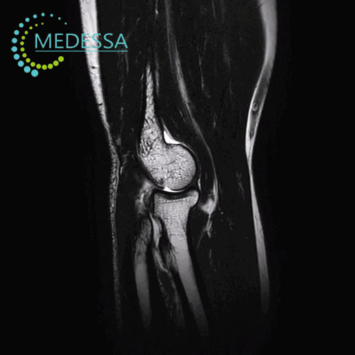
Reading the elbow as three connected articulations
The ulnohumeral articulation guides flexion and extension, the radiocapitellar articulation transmits load across the lateral side, and the proximal radioulnar articulation allows forearm rotation. MRI can assess the distal humerus, radial head, proximal ulna, their cartilage-covered surfaces, subchondral bone, marrow, capsule and synovial recesses within the selected field of view.
Because elbow cartilage is relatively thin, small defects may remain difficult to characterize. Joint incongruity, osteophytes, subchondral change or a suspected mineralized loose body may warrant radiographs or CT for complementary information.
Ligaments: visible damage versus functional instability
Medial ulnar collateral ligament
The medial collateral ligament complex, often termed the ulnar collateral ligament (UCL), is an important restraint to valgus loading. Its anterior bundle is particularly relevant in throwing and overhead activities. On MRI, a radiologist assesses fiber continuity, thickening, fluid-sensitive signal and surrounding edema, as well as changes at the humeral and ulnar attachments. A subtle partial defect may be less conspicuous on routine MRI than on selected direct MR arthrography. The degree of clinical instability cannot be established from ligament signal alone.
Lateral ligament supports
The lateral complex includes the radial collateral ligament (RCL), lateral ulnar collateral ligament (LUCL) and annular ligament. These structures support the radial head and contribute to resistance against posterolateral rotational instability. Scarring, discontinuity or associated soft-tissue injury may be visible, but the direction and functional significance of instability require clinical correlation.
Tendon attachments and patterns of pain
Epicondylar tendon origins
The common extensor origin at the lateral epicondyle may show thickening, altered internal signal or tearing in lateral epicondylopathy. Comparable findings at the common flexor-pronator origin are relevant to medial epicondylopathy. MRI can describe the extent of structural change and look for associated ligament or joint abnormalities; it cannot measure pain intensity from tendon appearance.
Distal biceps, brachialis and triceps
The distal biceps inserts on the radial tuberosity and is important for supination. Depending on coverage, MRI may depict a partial or complete tear, tendon retraction or bicipitoradial bursal fluid. The brachialis inserts on the proximal ulna and the triceps on the olecranon. Their attachments can demonstrate strain, tendinopathy, enthesopathic change or disruption. A suspected distal biceps lesion may require dedicated positioning or additional planes rather than assuming that every routine elbow series will depict its entire course equally well.
Bone marrow, cartilage and mechanical symptoms
Fluid-sensitive MRI images can reveal edema-like marrow signal following impact or repetitive loading, sometimes with an occult fracture line not evident on initial radiographs. Bone marrow edema is a nonspecific sign: its interpretation depends on the pattern, mechanism of injury and associated structural findings. CT may be needed to clarify cortical disruption or small osseous fragments.
At the capitellum, an osteochondral lesion or osteochondritis dissecans can affect the articular surface and underlying bone. MRI may demonstrate marrow change, cartilage abnormalities and findings that raise concern about fragment instability. It cannot determine fragment stability with certainty in every case. Mechanical catching or loss of extension may also prompt evaluation for intra-articular loose bodies or synovial disease.
Synovium, inflammation and focal lesions
Effusion, synovial thickening, erosive change and surrounding edema may occur in inflammatory arthropathy or infection. The imaging patterns overlap. MRI cannot identify the responsible microorganism or replace laboratory testing and aspiration when clinically indicated. Suspected joint infection requires prompt clinical assessment.
Within the scanned region, MRI may characterize a bursa, a postoperative abnormality or a mass and its relationship to adjacent structures. A mass’s exact histological identity cannot generally be determined by MRI alone; specialist evaluation and sometimes tissue sampling are necessary.
The ulnar nerve at the cubital tunnel
The ulnar nerve courses behind the medial epicondyle through the cubital tunnel. If adequately included, MRI can show nerve enlargement, abnormal signal or a nearby structural cause of compression. However, a normal-appearing nerve does not exclude intermittent entrapment or impaired nerve function. Examination, electrodiagnostic testing and sometimes dynamic ultrasound may be more informative for these questions.
Imaging planes and sequences: what each contributes
An elbow study combines planes, which determine the viewing direction, with MRI sequences, which emphasize different tissue characteristics. Axial views help follow tendon insertions, ligament relationships and the ulnar nerve in cross-section. Coronal views are useful for collateral ligaments and the radiocapitellar and ulnohumeral surfaces. Sagittal views provide information about flexor-extensor anatomy, the olecranon region, cartilage and joint recesses. Oblique or specially oriented images may be added for a specific tendon or ligament question.
- T1-weighted: outlines anatomy and normal fatty marrow; a change in marrow signal may draw attention to fracture-related or infiltrative abnormalities.
- PD-weighted and T2-weighted: contribute to assessment of cartilage, tendon and ligament structure, joint fluid and surrounding tissues, according to their acquisition settings.
- PD FS and T2 FS: suppress fat so edema-like signal, fluid and many traumatic or inflammatory abnormalities are easier to recognize.
- STIR: provides another fluid-sensitive method of highlighting marrow and soft-tissue edema.
The actual set of planes and sequences is chosen for the clinical question, patient positioning and technical circumstances. These examples describe imaging principles, not a promise that every sequence is performed in every examination.
Intravenous contrast, phases and direct MR arthrography
Routine elbow MRI commonly does not require intravenous gadolinium. Selected questions involving a mass, infection or active synovial inflammation may justify contrast, with T1-weighted fat-suppressed images before and after administration as appropriate. Dynamic post-contrast imaging is reserved for specific indications; it is not an obligatory phase of elbow MRI.
T1, T2, PD and STIR are sequences, not contrast phases. Direct MR arthrography is a separate technique in which diluted contrast is introduced into the joint under appropriate guidance. It may be useful for selected subtle intra-articular or ligament abnormalities and should not be confused with intravenous contrast-enhanced MRI.
Preparation, implant screening and image quality
Before scanning, patients should disclose implanted devices, previous surgery, retained metallic fragments, pregnancy or possible pregnancy, claustrophobia and difficulty remaining still. Devices are assessed using their exact documentation and MR Safe, MR Conditional or MR Unsafe status. MR Conditional conditions must be followed; the metal’s name alone is not sufficient to establish suitability.
If contrast is proposed, relevant medical history, previous reactions and renal function when indicated are reviewed according to safety procedures. Motion can obscure small tendon or ligament abnormalities. Metal near the elbow can produce artifacts, and limited field of view or spatial resolution can prevent reliable assessment of structures beyond the target area.
When additional imaging or clinical testing matters
Structural MRI findings do not necessarily identify the source of pain or quantify functional instability. Mild tendon signal changes may not correlate with symptoms; a subtle ligament defect or tiny loose body can be difficult to resolve. Radiographs help with initial trauma, alignment and mineralized abnormalities; CT can refine fracture or ossified-fragment assessment; ultrasound can assess selected superficial structures and dynamic abnormalities. Nerve-function tests, laboratory investigations or biopsy may be appropriate for specific clinical questions.
Related MRI examinations
- MRI of the Shoulder Joint — when symptoms are centered at the shoulder rather than the elbow.
- MRI of the Wrist Joint and Proximal Hand — for a separate wrist-region question.
- MRI of the Distal Hand — for symptoms localized to the hand or fingers.
Frequently asked questions about elbow MRI
Why does the referral need to identify the painful side of the elbow?
Medial, lateral, posterior and anterior symptoms suggest different structures. Describing the site and suspected problem helps determine which tendon, ligament or joint compartment needs particular attention.
Can elbow MRI show whether a ligament is stretched rather than torn?
MRI may show thickening, altered signal or fiber disruption, but it cannot reliably measure functional laxity. Examination and injury history are needed to interpret whether the ligament is clinically unstable.
Do I need MRI for ordinary tennis elbow?
Not necessarily. Many cases are evaluated clinically. Imaging may be considered when symptoms are atypical, persistent or raise concern about a significant tendon tear or another condition.
Can MRI explain why my elbow will not fully straighten?
It may identify joint fluid, an osteochondral lesion, synovial changes or a possible loose body. Examination and sometimes radiographs or CT help establish whether a mechanical obstruction is present.
Can a fracture be present if my first X-ray was normal?
Yes. Some occult injuries become apparent on MRI through a fracture line or surrounding marrow changes. Depending on the suspected fracture, CT or repeat radiographs may also be appropriate.
Will MRI tell me whether my ulnar nerve is working normally?
No. MRI evaluates visible nerve and surrounding tissue changes, not electrical function. Clinical testing and nerve-conduction studies may be needed, especially when symptoms come and go.
Will the scan cover pain extending to my wrist?
Not automatically. An elbow MRI is centered on the elbow and nearby attachments. A separate examination may be needed when the main concern lies farther along the forearm or at the wrist.
Is a contrast injection part of every elbow MRI?
No. Most routine structural questions are investigated without intravenous contrast. Contrast is considered for particular concerns such as a mass, infection or active synovial disease.
Can I have MRI after elbow fixation surgery?
It depends on the exact device and its documented MR safety conditions. Even when scanning is permitted, metal artifacts can limit the view, and a tailored examination may be required.
Does a normal MRI rule out the cause of persistent elbow pain?
No. Some dynamic instability, intermittent nerve compression and pain mechanisms may not be apparent on a static MRI. The result should be interpreted with symptoms and physical findings.
Material prepared and edited by: Andrii Bagirov
International medical sources and recommendations
- American College of Radiology — ACR Appropriateness Criteria: Chronic Elbow Pain.
- American College of Radiology — ACR Appropriateness Criteria: Acute Elbow and Forearm Pain.
- The elbow: review of anatomy and common collateral ligament complex pathology using MRI — peer-reviewed review article.
- American College of Radiology — Manual on Contrast Media.
- American College of Radiology — MR Safety resources.
Last updated: October 22, 2026.