
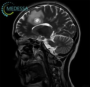
MRI of the Brain in Chornomorsk – Comprehensive Brain MRI on Philips Achieva 1.5 Tesla
Brain MRI is one of the most informative non-invasive imaging methods in modern neuroradiology. Magnetic resonance imaging of the brain allows detailed visualization of cerebral hemispheres, cortical and subcortical structures, white matter, gray matter, brainstem, cerebellum, pituitary region, ventricular system, cranial nerves, cerebrospinal fluid spaces, meninges, vascular structures, and surrounding soft tissues without ionizing radiation exposure.
At MEDESSA diagnostic center in Chornomorsk, brain MRI examinations are performed on the Philips Achieva 1.5 Tesla MRI system. High-quality 1.5T MRI remains the international diagnostic standard in neuroradiology and is widely used in leading European, American, and Asian medical centers. Modern 1.5 Tesla systems provide high soft tissue contrast, excellent visualization of white matter lesions, acute ischemic changes, tumors, demyelinating disease, inflammatory processes, and postoperative alterations while maintaining stable image quality and reduced susceptibility artifacts.
Brain MRI is critically important in the diagnosis of stroke, tumors, epilepsy, multiple sclerosis, traumatic brain injury, neurodegenerative disorders, encephalitis, vascular pathology, congenital abnormalities, hydrocephalus, and many other neurological conditions. MRI findings frequently influence further treatment planning, neurosurgical decisions, neurological therapy selection, oncologic staging, rehabilitation planning, and long-term patient monitoring.
Why 1.5 Tesla Brain MRI Remains the Clinical Standard
The Philips Achieva 1.5 Tesla MRI scanner used in MEDESSA Chornomorsk provides high diagnostic accuracy for routine and advanced brain imaging protocols. Contrary to common misconceptions, properly optimized 1.5T MRI protocols remain highly effective for the vast majority of neurological indications and are extensively used worldwide.
Advantages of modern 1.5 Tesla brain MRI include:
- High diagnostic reliability for acute and chronic neurological disorders;
- Excellent visualization of white matter lesions and demyelination;
- Accurate detection of acute ischemic stroke using DWI sequences;
- Reduced magnetic susceptibility artifacts compared to ultra-high-field systems in certain regions;
- Stable image quality for postoperative follow-up;
- Reliable contrast-enhanced imaging for tumors and inflammatory disease;
- Better compatibility with many MRI-conditional implants;
- High-quality MR angiography and cranial nerve assessment;
- Comprehensive neuroradiology protocols suitable for routine and complex neurological diagnostics.
What Brain MRI Shows
MRI of the brain allows detailed assessment of intracranial anatomy and pathological processes affecting the central nervous system. Depending on the clinical indication, MRI can detect both common and rare neurological diseases.
Brain MRI visualizes:
- Cerebral cortex and subcortical structures;
- White matter tracts;
- Basal ganglia;
- Thalami;
- Corpus callosum;
- Brainstem;
- Cerebellum;
- Pituitary gland and sellar region;
- Optic pathways;
- Internal auditory canals;
- Ventricular system;
- Cerebrospinal fluid spaces;
- Meninges;
- Intracranial arteries and veins;
- Cranial nerves;
- Orbital structures;
- Skull base and adjacent soft tissues.
Which MRI Sequences Are Used During Brain MRI
Modern brain MRI protocols include multiple MRI pulse sequences, each providing unique diagnostic information. Combined interpretation of these sequences significantly improves diagnostic accuracy.
T1-Weighted Imaging (T1)
T1-weighted images provide detailed anatomical visualization of brain structures. T1 sequences help assess normal anatomy, chronic hemorrhage, fat-containing lesions, postoperative changes, brain atrophy, and tissue morphology. After gadolinium administration, T1 imaging becomes essential for detecting contrast enhancement associated with tumors, inflammation, blood-brain barrier disruption, and active demyelinating lesions.
T2-Weighted Imaging (T2)
T2-weighted sequences are highly sensitive to increased water content and edema. They help identify ischemia, gliosis, inflammatory processes, tumors, cystic lesions, hydrocephalus, and white matter abnormalities. Many pathological processes appear hyperintense on T2 imaging.
FLAIR (Fluid-Attenuated Inversion Recovery)
FLAIR suppresses cerebrospinal fluid signal, significantly improving detection of periventricular and cortical lesions. This sequence is particularly important in multiple sclerosis, chronic ischemic disease, encephalitis, traumatic brain injury, and leptomeningeal pathology. FLAIR is one of the key sequences for detecting demyelinating plaques.
DWI (Diffusion-Weighted Imaging)
DWI is critically important in emergency neuroradiology and acute stroke diagnosis. Diffusion restriction may appear within minutes after ischemic injury. DWI is also valuable for detecting abscesses, highly cellular tumors, Creutzfeldt-Jakob disease, encephalitis, and some epileptic changes.
ADC (Apparent Diffusion Coefficient)
ADC maps complement DWI and help differentiate true restricted diffusion from T2 shine-through effects. Combined DWI/ADC interpretation improves accuracy in stroke characterization, tumor cellularity assessment, and differentiation between cytotoxic and vasogenic edema.
SWI (Susceptibility-Weighted Imaging)
SWI is highly sensitive to blood products, microhemorrhages, calcifications, venous structures, cavernomas, traumatic axonal injury, and hemorrhagic transformation of stroke. SWI is especially useful in neurovascular pathology and traumatic brain injury evaluation.
T1 with Contrast Enhancement (T1+C)
Post-contrast T1 imaging is essential for evaluating tumors, metastases, inflammatory disease, postoperative changes, abscesses, meningitis, active demyelinating lesions, and blood-brain barrier disruption.
TOF MRA (Time-of-Flight MR Angiography)
TOF MRA allows non-contrast visualization of intracranial arteries. This technique helps assess aneurysms, arterial stenosis, vascular malformations, vessel occlusions, congenital vascular variants, and cerebrovascular disease.
MRI of the Brain With Contrast Enhancement
Contrast-enhanced brain MRI significantly expands diagnostic capabilities in neuroradiology. Intravenous gadolinium-based contrast agents improve visualization of pathological tissue vascularity and blood-brain barrier disruption.
When Contrast Is Needed
Contrast-enhanced MRI may be recommended in cases of:
- Suspected brain tumors;
- Metastatic disease;
- Pituitary pathology;
- Multiple sclerosis activity assessment;
- Inflammatory and infectious disease;
- Meningitis and encephalitis;
- Postoperative follow-up;
- Evaluation of residual or recurrent tumors;
- Cranial nerve pathology;
- Unclear focal lesions on non-contrast MRI.
Gadolinium-Based Contrast Agents
Modern gadolinium contrast agents used in MRI are generally considered safe when administered appropriately. They do not contain iodine and differ fundamentally from CT contrast agents. Severe allergic reactions are rare.
Brain Tumors and Metastases
Contrast-enhanced MRI is one of the most important imaging methods for evaluation of primary brain tumors and intracranial metastases. Contrast enhancement patterns help assess tumor vascularity, necrosis, blood-brain barrier disruption, postoperative residual disease, and treatment response.
Multiple Sclerosis
Active demyelinating lesions may enhance after gadolinium administration. Contrast enhancement helps differentiate active inflammatory plaques from chronic inactive lesions and plays an important role in disease activity monitoring.
Inflammatory and Infectious Disorders
Contrast MRI improves detection of meningitis, encephalitis, abscesses, vasculitis, pachymeningitis, neurosarcoidosis, and autoimmune inflammatory disease affecting the central nervous system.
Postoperative MRI Follow-Up
Contrast-enhanced MRI is frequently used after neurosurgical interventions to evaluate postoperative anatomy, differentiate scar tissue from recurrent tumor, and monitor complications.
Contrast Safety and Kidney Function
Although gadolinium-based contrast agents are generally safe, kidney function assessment may be required in patients with severe renal impairment. In patients with advanced kidney failure, special precautions are necessary because of the rare risk of nephrogenic systemic fibrosis.
Indications for Brain MRI
- Chronic or severe headache;
- Migraine with neurological symptoms;
- Sudden neurological deficit;
- Acute stroke suspicion;
- Dizziness and balance disorders;
- Seizures and epilepsy;
- Visual disturbances;
- Memory impairment;
- Cognitive decline;
- Suspected multiple sclerosis;
- Brain tumor suspicion;
- Traumatic brain injury;
- Hearing disturbances;
- Pituitary disorders;
- Cranial nerve pathology;
- Hydrocephalus;
- Inflammatory neurological disease;
- Follow-up after neurosurgery;
- Monitoring of known intracranial pathology.
Which Symptoms May Indicate Brain Pathology
Neurological symptoms may reflect structural, vascular, inflammatory, neoplastic, degenerative, or demyelinating disease affecting the brain.
- Persistent or progressive headache;
- New-onset severe headache after age 50;
- Sudden thunderclap headache;
- Focal weakness or numbness;
- Speech disturbances;
- Facial asymmetry;
- Seizures;
- Visual field defects;
- Double vision;
- Memory decline;
- Confusion;
- Gait instability;
- Loss of consciousness;
- Behavioral changes;
- Persistent dizziness;
- Tremor or movement disorders.
Headache and Neurological Red Flags
Although many headaches are benign, certain clinical features may require urgent neuroimaging evaluation.
Important red flags include:
- Sudden “worst headache of life”;
- Headache with focal neurological deficit;
- Headache with seizures;
- Progressively worsening headache;
- Headache associated with fever or neck stiffness;
- Headache after head trauma;
- Headache with altered consciousness;
- Headache in patients with cancer history;
- New neurological symptoms;
- Immunosuppression or systemic inflammatory disease.
MRI of the Brain in Multiple Sclerosis
MRI plays a central role in the diagnosis and monitoring of multiple sclerosis (MS). Demyelinating plaques typically involve periventricular white matter, juxtacortical regions, infratentorial structures, corpus callosum, and spinal cord.
Demyelination
Multiple sclerosis causes inflammatory demyelination of central nervous system white matter tracts. MRI allows detection of both symptomatic and asymptomatic lesions.
FLAIR Lesions
FLAIR imaging is especially sensitive for identifying periventricular and juxtacortical plaques characteristic of MS.
Active Lesions
Gadolinium-enhancing lesions may indicate active inflammation and ongoing blood-brain barrier disruption.
McDonald Criteria
Modern MS diagnosis relies heavily on MRI findings according to the McDonald criteria, including dissemination in space and dissemination in time.
Diseases Most Commonly Detected on Brain MRI
Stroke
MRI is highly sensitive for acute ischemic stroke detection using DWI and ADC sequences. MRI also evaluates chronic ischemic disease, lacunar infarcts, hemorrhagic transformation, and microvascular pathology.
Brain Tumors
MRI detects gliomas, meningiomas, pituitary adenomas, vestibular schwannomas, metastases, lymphomas, and many other intracranial neoplasms.
Multiple Sclerosis
MRI is the primary imaging method for diagnosing and monitoring demyelinating disease activity.
Encephalitis
MRI can identify inflammatory changes affecting temporal lobes, limbic structures, cortical regions, and white matter in infectious or autoimmune encephalitis.
Epilepsy
Dedicated epilepsy MRI protocols may reveal hippocampal sclerosis, cortical dysplasia, developmental abnormalities, tumors, vascular malformations, and post-traumatic epileptogenic lesions.
Dementia and Neurodegeneration
MRI helps evaluate cortical atrophy patterns, hippocampal volume loss, vascular dementia changes, normal pressure hydrocephalus, and neurodegenerative disorders.
Aneurysms and Vascular Pathology
MR angiography helps detect aneurysms, arterial stenosis, vascular malformations, congenital variants, and vessel occlusions.
When Brain MRI Is Needed Urgently
Emergency MRI or urgent neuroimaging evaluation may be necessary in potentially life-threatening neurological conditions.
- Acute ischemic stroke;
- Sudden focal neurological deficit;
- Status epilepticus;
- New-onset seizures;
- Acute consciousness impairment;
- Suspected encephalitis;
- Rapid neurological deterioration;
- Acute severe headache with neurological symptoms;
- Suspected intracranial hemorrhage;
- Post-traumatic neurological decline.
Contraindications to Brain MRI
Absolute Contraindications
- Certain non-MRI-compatible cardiac pacemakers;
- Some ferromagnetic intracranial aneurysm clips;
- Unsafe implanted electronic devices;
- Certain metallic foreign bodies.
Relative Contraindications
- Severe claustrophobia;
- Inability to remain still;
- First trimester pregnancy in selected cases;
- Some MRI-conditional implants requiring protocol verification.
Cardiac Pacemakers and Implants
Many modern implants are MRI-conditional, but compatibility must always be verified before scanning.
Claustrophobia
Patients with claustrophobia may require psychological support, mild sedation, or adapted examination strategies.
Metallic Foreign Bodies
Metal fragments, especially intraorbital metallic foreign bodies, require careful safety assessment before MRI.
Preparation for Brain MRI
Metallic Objects
Patients should remove metallic accessories, watches, jewelry, hearing aids, removable dental appliances, and electronic devices before scanning.
Medical Documentation
Previous MRI, CT, PET, ultrasound, surgical reports, oncology records, and neurological evaluations may significantly improve diagnostic interpretation.
Creatinine Testing Before Contrast MRI
Patients with kidney disease, diabetes, or advanced age may require renal function assessment before gadolinium administration.
Sedation
In selected patients with severe anxiety, claustrophobia, or inability to remain still, sedation may be considered.
Children
Pediatric brain MRI sometimes requires sedation depending on the child’s age and ability to cooperate during scanning.
Comparison Between Brain MRI and Brain CT
| Feature | Brain MRI | Brain CT |
|---|---|---|
| Radiation exposure | No ionizing radiation | Uses ionizing radiation |
| Soft tissue visualization | Excellent | Limited compared to MRI |
| Acute stroke sensitivity | Very high with DWI | Good for hemorrhage |
| Brain tumors | Highly sensitive | Less detailed |
| Multiple sclerosis | Primary imaging method | Poor sensitivity |
| Bone assessment | Limited | Excellent |
| Microhemorrhage detection | Excellent with SWI | Limited |
| Emergency speed | Longer examination | Faster acquisition |
What Brain MRI May Not Show
Although MRI is an extremely powerful diagnostic tool, it has limitations and should not be viewed as an “all-powerful” examination. Some neurological symptoms may occur without detectable structural abnormalities on MRI.
Brain MRI may have limited sensitivity in:
- Very early microscopic pathological changes;
- Certain metabolic disorders;
- Functional neurological disorders;
- Some psychiatric conditions;
- Microscopic axonal injury below imaging resolution;
- Early neurodegenerative disease without structural changes;
- Some migraine-related disorders;
- Electrophysiological abnormalities without anatomical correlates.
MRI findings should always be interpreted together with neurological examination, laboratory testing, patient history, and other diagnostic methods.
Related MRI Examinations
Frequently Asked Questions (FAQ)
What does brain MRI show?
Brain MRI visualizes brain tissue, white matter, gray matter, ventricles, cranial nerves, meninges, blood vessels, pituitary structures, cerebellum, and brainstem. It helps detect stroke, tumors, multiple sclerosis, inflammation, vascular abnormalities, traumatic injury, and neurodegenerative changes.
Can MRI detect a stroke?
Yes. MRI with diffusion-weighted imaging is highly sensitive for detecting acute ischemic stroke, including very early ischemic changes that may not yet be visible on CT.
Is contrast always necessary for brain MRI?
No. Contrast is used selectively depending on the clinical indication, suspected tumor, inflammation, demyelinating disease, infection, postoperative follow-up, or unclear findings on non-contrast imaging.
How long does brain MRI take?
Standard brain MRI usually takes approximately 20–40 minutes depending on protocol complexity and whether contrast enhancement or angiography is required.
Can MRI detect multiple sclerosis?
MRI is the primary imaging method for diagnosing and monitoring multiple sclerosis. FLAIR and contrast-enhanced sequences are especially important for detecting demyelinating lesions.
Can MRI detect brain aneurysms?
MR angiography can help visualize aneurysms, arterial stenosis, vascular malformations, and intracranial vessel abnormalities.
Is brain MRI safe?
MRI does not use ionizing radiation and is generally considered safe when MRI safety recommendations are followed appropriately.
Can MRI detect epilepsy-related abnormalities?
Yes. Specialized epilepsy MRI protocols may reveal hippocampal sclerosis, cortical dysplasia, developmental abnormalities, tumors, and other epileptogenic lesions.
Can MRI detect dementia?
MRI may show cortical atrophy, hippocampal volume loss, vascular changes, and other structural findings associated with neurodegenerative disease.
What is the difference between MRI and CT of the brain?
MRI provides superior soft tissue visualization without radiation exposure, while CT is faster and particularly useful for acute hemorrhage and bone assessment.
Can MRI miss a brain disorder?
Yes. Some neurological disorders may not produce visible structural changes on MRI, especially during very early stages or in primarily functional conditions.