
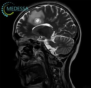
Brain MRI (Magnetic Resonance Imaging of the Brain)
Brain MRI is a high-resolution, non-invasive neuroimaging examination used to evaluate the brain, meninges, cerebrospinal fluid spaces, intracranial vessels, cranial nerves, pituitary region, and surrounding anatomical structures. MRI does not use ionizing radiation and provides excellent soft-tissue contrast, making it one of the most informative methods for detecting structural, vascular, inflammatory, demyelinating, traumatic, neoplastic, infectious, and degenerative diseases of the central nervous system.
Magnetic Resonance Imaging of the brain is especially valuable when symptoms are non-specific, when early disease is suspected, or when detailed anatomical assessment is required for diagnosis, treatment planning, neurosurgical preparation, radiotherapy planning, or follow-up after therapy. Depending on the clinical question, a brain MRI protocol may include conventional anatomical sequences, diffusion imaging, susceptibility-sensitive sequences, contrast-enhanced imaging, MR angiography, MR venography, perfusion MRI, or MR spectroscopy.
What Structures Are Evaluated on Brain MRI?
A properly performed MRI of the brain allows detailed visualization of the major intracranial structures and helps assess their morphology, signal characteristics, symmetry, volume, and relationship to surrounding tissues.
- cerebral hemispheres, including the frontal, parietal, temporal, and occipital lobes;
- cerebral cortex, subcortical white matter, deep white matter, and corpus callosum;
- basal ganglia, thalamus, internal capsule, and deep gray matter nuclei;
- brainstem, including the midbrain, pons, and medulla oblongata;
- cerebellum, cerebellar hemispheres, vermis, and cerebellar peduncles;
- ventricular system, including the lateral ventricles, third ventricle, cerebral aqueduct, and fourth ventricle;
- subarachnoid spaces, cisterns, and cerebrospinal fluid pathways;
- meninges, dural venous sinuses, and extra-axial spaces;
- pituitary gland, sella turcica, suprasellar region, and hypothalamus;
- optic nerves, optic chiasm, optic tracts, and visual pathways;
- cranial nerves, especially when dedicated high-resolution protocols are used;
- major intracranial arteries and veins when MR angiography or venography is added;
- skull base, cerebellopontine angles, internal auditory canals, and adjacent soft tissues.
What Can Brain MRI Detect?
Brain MRI can identify a wide spectrum of abnormalities, from frequent neurological diseases to rare and complex intracranial conditions. It is used not only to detect pathology, but also to define its extent, relationship to critical structures, biological behavior, complications, and response to treatment.
- ischemic stroke, acute infarction, chronic ischemic changes, lacunar infarcts, and post-stroke gliosis;
- intracranial hemorrhage, microbleeds, cavernomas, hemorrhagic transformation, and chronic hemosiderin deposits;
- primary brain tumors, including gliomas, meningiomas, ependymomas, medulloblastomas, and other neoplasms;
- brain metastases, leptomeningeal spread, and treatment-related tumor changes;
- demyelinating diseases, including multiple sclerosis, acute disseminated encephalomyelitis, MOG-associated disease, and neuromyelitis optica spectrum disorders;
- inflammatory and infectious diseases such as encephalitis, meningitis, cerebritis, brain abscess, and opportunistic infections;
- epileptogenic lesions, including hippocampal sclerosis, focal cortical dysplasia, low-grade tumors, vascular malformations, and post-traumatic gliosis;
- congenital and developmental brain malformations, including Chiari malformation, cortical malformations, agenesis or dysgenesis of the corpus callosum, and posterior fossa anomalies;
- hydrocephalus, CSF circulation disorders, intracranial hypotension, and intracranial hypertension-related changes;
- neurodegenerative disorders with structural brain atrophy patterns, including dementia-related changes;
- traumatic brain injury, contusions, diffuse axonal injury, subdural collections, and chronic post-traumatic changes;
- vascular abnormalities such as aneurysms, arteriovenous malformations, dural arteriovenous fistulas, venous thrombosis, and vasculopathy when vascular MRI techniques are included.
Common Indications for Brain MRI
- persistent, progressive, unusual, or unexplained headache;
- dizziness, imbalance, vertigo, coordination problems, or gait disturbance;
- suspected stroke, transient ischemic attack, or chronic cerebrovascular disease;
- seizures, epilepsy, loss of consciousness, or unexplained paroxysmal neurological episodes;
- visual disturbances, double vision, optic neuritis, or suspected pathology of the visual pathways;
- hearing loss, tinnitus, facial numbness, facial pain, or cranial nerve symptoms;
- memory impairment, cognitive decline, behavioral changes, or suspected dementia;
- suspected multiple sclerosis or another demyelinating disease;
- suspected brain tumor, metastasis, pituitary lesion, or intracranial mass effect;
- evaluation after traumatic brain injury;
- fever, altered mental status, or suspicion of encephalitis, meningitis, or brain abscess;
- postoperative follow-up after neurosurgery, radiation therapy, chemotherapy, or targeted treatment;
- monitoring of known neurological disease over time.
Which MRI Sequences Are Used in Brain MRI?
A brain MRI examination is not a single image but a combination of multiple MRI sequences. Each sequence highlights different tissue characteristics and helps answer a specific diagnostic question. The final protocol is selected according to the patient’s symptoms, clinical history, suspected diagnosis, and whether contrast enhancement is required.
T1-Weighted Imaging
T1-weighted images provide detailed anatomical information and are useful for evaluating normal brain morphology, cortical and subcortical structures, fat-containing lesions, subacute hemorrhage, and the relationship between a lesion and adjacent anatomy. T1 images are also the baseline for comparison with post-contrast T1-weighted sequences.
T2-Weighted Imaging
T2-weighted imaging is highly sensitive to increased water content. It helps detect edema, inflammation, gliosis, chronic infarcts, cystic lesions, demyelinating plaques, tumors, and many structural abnormalities. Pathological processes with increased fluid content often appear hyperintense on T2-weighted images.
FLAIR (Fluid-Attenuated Inversion Recovery)
FLAIR suppresses the signal from free cerebrospinal fluid, making lesions near the ventricles and cortical surfaces more conspicuous. It is particularly important in the assessment of multiple sclerosis, chronic ischemic changes, gliosis, encephalitis, meningitis-related abnormalities, subarachnoid pathology, and cortical or juxtacortical lesions.
DWI (Diffusion-Weighted Imaging)
DWI evaluates the movement of water molecules within tissues. It is essential for detecting acute ischemic stroke, often within minutes to hours after symptom onset. DWI is also useful in identifying abscesses, highly cellular tumors, epidermoid cysts, some encephalitic lesions, and areas of cytotoxic edema.
ADC Map (Apparent Diffusion Coefficient)
ADC maps are interpreted together with DWI to distinguish true restricted diffusion from T2 shine-through. Reduced ADC values support true diffusion restriction, which is typical for acute infarction, pus in abscesses, and some highly cellular tumors. ADC is important for accurate interpretation of acute and subacute brain lesions.
SWI (Susceptibility-Weighted Imaging)
SWI is highly sensitive to magnetic susceptibility effects caused by blood products, calcifications, venous structures, and mineral deposition. It is useful for detecting microbleeds, cavernous malformations, traumatic diffuse axonal injury, hemorrhagic lesions, venous thrombosis signs, and small foci of hemosiderin that may be invisible on routine sequences.
T1+C (Contrast-Enhanced T1-Weighted Imaging)
Post-contrast T1-weighted imaging is performed after intravenous administration of a gadolinium-based contrast agent. It helps detect abnormal blood-brain barrier disruption, tumor enhancement, metastases, active demyelinating plaques, meningeal enhancement, abscess capsules, inflammatory lesions, and postoperative residual or recurrent disease.
TOF MRA (Time-of-Flight MR Angiography)
TOF MRA is a non-contrast MR angiographic technique used to visualize arterial blood flow. It is commonly used to assess intracranial arteries, detect aneurysms, stenoses, occlusions, vascular malformations, and arterial variants. If vascular pathology is suspected, TOF MRA may be added to a standard brain MRI protocol.
Brain MRI With Contrast
Brain MRI with contrast is performed when it is necessary to evaluate abnormal enhancement patterns, blood-brain barrier disruption, tumor vascularity, inflammatory activity, meningeal involvement, or postoperative changes. Contrast-enhanced MRI does not replace non-contrast sequences; it complements them and provides additional diagnostic information when clinically indicated.
When Is Contrast Needed for Brain MRI?
- suspected primary brain tumor or tumor recurrence;
- search for brain metastases, especially small or multiple lesions;
- evaluation of meningeal or leptomeningeal disease;
- suspected active multiple sclerosis lesions;
- suspected encephalitis, meningitis, cerebritis, or brain abscess;
- assessment of pituitary tumors and sellar region pathology;
- postoperative follow-up after tumor resection or neurosurgical intervention;
- differentiation between scar tissue, radiation necrosis, residual tumor, and recurrent disease;
- evaluation of inflammatory, autoimmune, granulomatous, or infectious CNS disease.
Gadolinium-Based Contrast Agents
Most MRI contrast agents used for brain imaging contain gadolinium in a chelated form. Gadolinium-based contrast agents help demonstrate areas where the blood-brain barrier is disrupted or where abnormal vascularity is present. Enhancement patterns can provide important diagnostic clues, such as ring enhancement in abscess or necrotic tumors, homogeneous enhancement in some meningiomas, nodular enhancement in metastases, or linear meningeal enhancement in inflammatory or neoplastic meningeal disease.
Contrast MRI in Brain Tumors and Metastases
Contrast-enhanced brain MRI is highly important in neuro-oncology. It helps identify tumor margins, necrotic components, edema, multifocal lesions, leptomeningeal spread, and postoperative residual enhancement. In patients with known cancer, contrast MRI is often the preferred method for detecting brain metastases, especially small lesions that may be difficult to see on non-contrast imaging.
Contrast MRI in Multiple Sclerosis
In multiple sclerosis, gadolinium enhancement may indicate active inflammatory demyelination. Enhancing lesions can help demonstrate dissemination in time when interpreted together with non-enhancing lesions, clinical data, and established diagnostic criteria. Contrast is especially useful when assessing suspected disease activity, relapse, or treatment response.
Safety of MRI Contrast and Kidney Function
Gadolinium-based contrast agents are generally well tolerated, but they must be used appropriately. Before contrast administration, patients may be asked about kidney disease, previous contrast reactions, pregnancy, and relevant medical history. In patients with severe renal impairment or acute kidney injury, the decision to administer gadolinium requires careful risk-benefit assessment, because impaired renal clearance increases the risk of rare contrast-related complications such as nephrogenic systemic fibrosis.
If contrast is planned, serum creatinine or estimated glomerular filtration rate (eGFR) may be required, especially in patients with known kidney disease, diabetes, advanced age, hypertension, previous kidney surgery, or other risk factors for renal dysfunction. The radiologist and referring physician determine whether contrast is necessary and safe in each individual case.
Contraindications to Brain MRI
MRI safety screening is mandatory before every examination. Contraindications depend on the type of implant, material composition, device model, implantation date, magnetic field compatibility, and the patient’s clinical condition. Many modern implants are MR Conditional, meaning MRI may be possible only under specific technical conditions.
Absolute Contraindications
- non-MRI-compatible cardiac pacemakers, implantable cardioverter-defibrillators, or neurostimulators;
- ferromagnetic intracranial aneurysm clips or implants with unknown MRI safety status;
- certain older cochlear implants or electronic implants not approved for MRI;
- metallic foreign bodies in critical locations, especially intraorbital metallic fragments;
- any implanted device that cannot be verified as MR Safe or MR Conditional when risk is significant.
Relative Contraindications and Special Situations
- claustrophobia or severe anxiety requiring preparation, support, or sedation;
- pregnancy, especially during the first trimester, when MRI is performed only when clinically justified;
- renal insufficiency when contrast-enhanced MRI is planned;
- recent surgery or recently implanted medical devices;
- metallic implants, orthopedic hardware, vascular stents, shunts, valves, or surgical clips requiring safety verification;
- inability to remain still due to pain, tremor, involuntary movements, or altered mental status;
- children or patients who may require sedation to obtain diagnostic-quality images.
Metallic Fragments and Eye Safety
Patients with a history of metalwork, welding, penetrating trauma, military injury, shrapnel, or suspected metallic foreign bodies must inform the medical team before MRI. Metallic fragments near the eyes or brain may be dangerous in a magnetic field. If needed, additional screening such as X-ray or CT may be performed before MRI.
Preparation for Brain MRI
Most non-contrast brain MRI examinations do not require complex preparation. However, correct preparation improves safety, image quality, and diagnostic accuracy.
- Remove metallic items before entering the MRI room, including jewelry, watches, hairpins, piercings, removable dental appliances, glasses, bank cards, keys, and electronic devices.
- Bring a referral, clinical summary, neurological examination notes, laboratory data, and relevant medical documentation when available.
- Bring previous MRI, CT, PET/CT, angiography, or ultrasound results for comparison, especially if the examination is performed for tumor follow-up, multiple sclerosis, stroke, epilepsy, or postoperative control.
- Inform the staff about pacemakers, implants, surgical clips, stents, shunts, prostheses, cochlear implants, neurostimulators, infusion pumps, or any implanted electronic device.
- If contrast MRI is planned, serum creatinine or eGFR may be required to assess kidney function.
- Tell the medical team about pregnancy, breastfeeding, kidney disease, previous allergic-like reactions to contrast agents, or severe chronic illness.
- If claustrophobia is present, discuss possible preparation options in advance.
- Children may require age-appropriate explanation, immobilization techniques, or sedation depending on age, clinical condition, and ability to remain still.
Brain MRI in Multiple Sclerosis
MRI is central to the diagnosis and monitoring of multiple sclerosis. It helps detect demyelinating plaques in characteristic locations, assess lesion burden, identify active inflammation, and evaluate disease progression over time. MRI findings are interpreted together with clinical symptoms, neurological examination, laboratory data, and diagnostic criteria.
Demyelination and Typical Lesion Locations
In multiple sclerosis, MRI may show demyelinating lesions in the periventricular white matter, juxtacortical and cortical regions, infratentorial structures, brainstem, cerebellum, optic nerves, and spinal cord. FLAIR is particularly useful for identifying periventricular and juxtacortical plaques.
FLAIR Lesions
FLAIR hyperintense lesions are one of the key MRI features in suspected demyelinating disease. Their number, morphology, distribution, and relationship to ventricles and cortex are important for distinguishing multiple sclerosis from migraine-related white matter lesions, small vessel ischemic disease, vasculitis, infection, and other mimics.
Active Lesions and Contrast Enhancement
Active inflammatory plaques may enhance after gadolinium administration. The presence of both enhancing and non-enhancing lesions can support dissemination in time, depending on the clinical context and diagnostic criteria.
McDonald Criteria and MRI
The McDonald criteria use clinical and MRI evidence to support the diagnosis of multiple sclerosis. MRI helps demonstrate dissemination in space and dissemination in time. However, MRI findings alone are not sufficient in every case; diagnosis requires correlation with symptoms, neurological examination, cerebrospinal fluid analysis when indicated, and exclusion of alternative diagnoses.
Brain MRI in Headache Evaluation
Headache is one of the most common reasons for neurological consultation. Many headaches are primary headache disorders, such as migraine or tension-type headache, and may not be associated with structural abnormalities. However, MRI is important when headache has atypical features, neurological symptoms, sudden onset, progressive course, or clinical red flags suggesting secondary intracranial pathology.
Red Flags That May Require Brain MRI
- sudden severe “thunderclap” headache;
- new or progressive headache after the age of 50;
- headache with weakness, numbness, speech disturbance, visual loss, double vision, or coordination problems;
- headache associated with seizures, confusion, fainting, or altered consciousness;
- headache that worsens with coughing, sneezing, straining, or changes in body position;
- headache that wakes the patient from sleep or is worse in the morning;
- headache in a patient with known cancer, immunosuppression, or systemic infection;
- new headache during pregnancy or postpartum period;
- headache after head trauma;
- headache accompanied by fever, neck stiffness, or signs of meningitis.
When Is Brain MRI Needed Urgently?
Urgent neuroimaging may be required when symptoms suggest an acute neurological condition. In emergency settings, CT may be performed first because it is fast and widely available, especially for suspected acute hemorrhage. MRI is particularly valuable for detecting early ischemia, posterior fossa stroke, encephalitis, diffuse axonal injury, and complex neurological emergencies.
- suspected acute ischemic stroke or transient ischemic attack;
- new-onset seizures or recurrent unexplained seizures;
- acute neurological deficit, including weakness, numbness, facial asymmetry, speech disturbance, or visual loss;
- altered consciousness, confusion, coma, or unexplained mental status change;
- suspected encephalitis, meningitis, or brain abscess;
- severe sudden headache or suspected intracranial bleeding;
- rapidly progressive neurological symptoms;
- suspected cerebral venous sinus thrombosis;
- neurological deterioration after head trauma.
Most Common Diseases Detected by Brain MRI
Stroke and Cerebrovascular Disease
MRI is highly sensitive for detecting acute ischemic stroke using DWI and ADC maps. It also shows chronic ischemic lesions, lacunar infarcts, leukoaraiosis, post-stroke gliosis, microbleeds, and vascular-related brain damage.
Brain Tumors and Metastases
MRI helps detect intracranial tumors, define tumor location and extent, assess edema and mass effect, evaluate contrast enhancement, and support treatment planning. Contrast-enhanced MRI is especially important for detecting metastases and postoperative recurrence.
Multiple Sclerosis
MRI detects demyelinating plaques, evaluates lesion distribution, identifies active enhancing lesions, and monitors changes over time. It is one of the most important tools in confirming and following multiple sclerosis.
Encephalitis and Inflammatory Diseases
MRI can reveal inflammatory signal abnormalities in the temporal lobes, limbic system, cortex, white matter, brainstem, or cerebellum, depending on the underlying cause. It is useful in viral, autoimmune, and other inflammatory encephalitides.
Epilepsy-Related Brain Abnormalities
Dedicated epilepsy MRI protocols can detect hippocampal sclerosis, focal cortical dysplasia, developmental malformations, post-traumatic gliosis, vascular malformations, and low-grade tumors that may be associated with seizures.
Dementia and Neurodegenerative Disorders
Brain MRI can assess global and regional atrophy, vascular contributions to cognitive impairment, normal pressure hydrocephalus features, prior infarcts, microangiopathic changes, and other structural causes of cognitive decline.
Aneurysms and Vascular Malformations
Standard brain MRI may show indirect signs of vascular lesions, while MR angiography is used to assess intracranial arteries more directly. When aneurysm, stenosis, arteriovenous malformation, or vascular anomaly is suspected, MR angiography may be recommended.
Symptoms That May Indicate Brain Pathology
Brain pathology may present with neurological, cognitive, sensory, motor, behavioral, or systemic symptoms. MRI may be recommended when symptoms are persistent, progressive, unexplained, focal, or associated with neurological examination abnormalities.
- persistent headache or change in headache pattern;
- dizziness, imbalance, vertigo, or gait instability;
- weakness, numbness, tingling, or loss of coordination;
- speech difficulty, confusion, memory decline, or cognitive impairment;
- vision loss, double vision, visual field defects, or optic neuritis symptoms;
- hearing loss, tinnitus, facial pain, or facial numbness;
- seizures, fainting, or episodes of altered awareness;
- personality changes, behavioral changes, or unexplained psychiatric symptoms with neurological concern;
- hormonal disturbances suggesting pituitary or hypothalamic pathology;
- fever, neck stiffness, or signs of central nervous system infection;
- progressive neurological deterioration.
Brain MRI vs Brain CT
MRI and CT are complementary imaging methods. The best choice depends on the clinical situation, urgency, suspected diagnosis, contraindications, and availability. CT is often preferred in acute trauma and suspected acute bleeding because it is fast. MRI provides superior soft-tissue detail and is more sensitive for many brain diseases, especially early ischemia, demyelination, posterior fossa lesions, tumors, inflammation, and epilepsy-related abnormalities.
| Comparison | Brain MRI | Brain CT |
|---|---|---|
| Radiation exposure | No ionizing radiation | Uses ionizing radiation |
| Soft-tissue detail | Excellent soft-tissue contrast | Lower soft-tissue contrast compared with MRI |
| Acute ischemic stroke | Very sensitive with DWI and ADC | May be normal early after symptom onset |
| Acute hemorrhage | Can detect hemorrhage, especially with SWI/GRE | Often first-line in emergency settings |
| Brain tumors | Preferred for tumor characterization and treatment planning | Can detect mass effect and calcification, but less detailed |
| Multiple sclerosis | Preferred method for detecting demyelinating plaques | Limited diagnostic value for MS |
| Posterior fossa and brainstem | High diagnostic value | May be limited by bone artifacts |
| Trauma | Useful for diffuse axonal injury and subacute complications | Often first-line for acute trauma |
| Examination time | Usually longer | Usually faster |
| Implant limitations | Requires strict MRI safety screening | Fewer implant-related restrictions |
What Brain MRI May Not Show
Brain MRI is a highly informative diagnostic tool, but it is not “all-seeing” and should not be interpreted outside the clinical context. A normal MRI does not always exclude functional, metabolic, microscopic, genetic, early inflammatory, or intermittent neurological disorders.
- very early or subtle disease may be below the resolution of routine MRI;
- some forms of migraine, functional neurological symptoms, and many primary headache disorders may not produce visible structural changes;
- electrical seizure activity itself is not directly visualized by MRI and requires EEG correlation;
- some metabolic, toxic, autoimmune, or genetic disorders may require laboratory testing, CSF analysis, genetic testing, or specialized imaging;
- small vascular abnormalities may require dedicated MR angiography, CT angiography, or catheter angiography;
- microscopic tumor infiltration may extend beyond visible MRI abnormalities;
- image quality can be reduced by motion, dental hardware artifacts, implants, or inability to complete the examination;
- interpretation depends on the correct protocol, clinical information, comparison with previous imaging, and radiologist expertise.
How Brain MRI Helps Treatment Planning
Brain MRI provides information that directly influences clinical decision-making. It helps neurologists, neurosurgeons, oncologists, radiation oncologists, infectious disease specialists, and other physicians choose the most appropriate treatment strategy.
- defines tumor location, size, edema, mass effect, and relationship to eloquent brain areas;
- helps plan neurosurgical biopsy, tumor resection, stereotactic radiosurgery, or radiation therapy fields;
- monitors treatment response after surgery, chemotherapy, radiotherapy, immunotherapy, or targeted therapy;
- supports diagnosis and follow-up in multiple sclerosis and other demyelinating diseases;
- helps determine stroke age, infarct extent, hemorrhagic transformation, and chronic vascular burden;
- identifies complications such as abscess, hydrocephalus, venous thrombosis, or postoperative recurrence;
- provides baseline imaging for long-term comparison in chronic neurological disease.
Related MRI Examinations
Depending on symptoms and clinical suspicion, brain MRI may be complemented by more specialized MRI examinations:
- MR Angiography of Brain Vessels — for evaluation of intracranial arteries, aneurysms, stenosis, occlusion, and vascular malformations;
- MR Angiography of Neck Vessels — for assessment of carotid and vertebral arteries, stenosis, dissection, and cerebrovascular risk factors;
- Pituitary MRI — for detailed evaluation of the pituitary gland, pituitary adenomas, hormonal disorders, and sellar region pathology;
- Orbital MRI — for assessment of optic nerves, extraocular muscles, orbital masses, inflammation, and visual pathway pathology;
- MRI for Epilepsy — for detection of epileptogenic structural lesions using a dedicated epilepsy protocol;
- MR Spectroscopy — for metabolic assessment of brain lesions, tumor characterization, and differentiation of selected pathological processes.
Frequently Asked Questions (FAQ)
What does a brain MRI show?
Brain MRI shows the brain parenchyma, ventricular system, meninges, brainstem, cerebellum, pituitary region, cerebrospinal fluid spaces, cranial nerves, and many intracranial vascular structures. It can detect stroke, tumors, metastases, demyelination, inflammation, traumatic injury, hydrocephalus, hemorrhage, and vascular abnormalities.
Can MRI detect a stroke?
Yes. Brain MRI is especially sensitive for acute ischemic stroke when diffusion-weighted imaging (DWI) and ADC maps are used. It can also assess chronic ischemic changes, lacunar infarcts, hemorrhagic transformation, microbleeds, and consequences of previous cerebrovascular events.
Is contrast needed for brain MRI?
Contrast is not always required. It is used when there is suspicion of a tumor, metastases, active inflammation, multiple sclerosis activity, meningeal involvement, infection, postoperative changes, or unclear abnormalities on non-contrast MRI.
Can MRI show multiple sclerosis?
Yes. MRI is one of the main methods for detecting demyelinating lesions in multiple sclerosis. T2 and FLAIR sequences are especially important, while contrast-enhanced imaging helps identify active inflammatory lesions.
Can brain MRI find the cause of headache?
Brain MRI can detect structural causes of headache, including tumors, hydrocephalus, inflammation, post-traumatic changes, vascular abnormalities, Chiari malformation, or signs of intracranial hypertension. However, in primary migraine or tension-type headache, MRI may be normal.
Which is better: brain MRI or brain CT?
MRI provides better soft-tissue detail and is more informative for demyelination, tumors, encephalitis, epilepsy-related abnormalities, posterior fossa lesions, and early ischemia. CT is often used first in emergency settings to rapidly exclude acute hemorrhage or skull fracture.
Is brain MRI safe?
Brain MRI does not use ionizing radiation and is considered safe when MRI safety rules are followed. It is essential to inform the medical team about pacemakers, implants, metallic fragments, neurostimulators, cochlear implants, or any other potentially unsafe device.
How long does a brain MRI take?
The duration depends on the protocol. A standard brain MRI is usually shorter, while MRI with contrast, MR angiography, epilepsy protocol, spectroscopy, or additional specialized sequences may take longer.
Can children have a brain MRI?
Yes. Brain MRI can be performed in children when medically indicated. The main requirement is that the child remains still during scanning. In young children or patients unable to cooperate, sedation may be considered by the physician.
Should I bring previous imaging results?
Yes. Previous MRI, CT, hospital discharge summaries, neurological reports, and treatment records help the radiologist compare findings over time, evaluate disease activity, and distinguish old changes from new pathology.
Can brain MRI be normal even when symptoms are present?
Yes. Some functional, metabolic, electrical, microscopic, or early pathological conditions may not produce visible structural changes on routine MRI. In such cases, a physician may recommend additional tests or a specialized MRI protocol.
Can MRI detect a brain aneurysm?
Sometimes large aneurysms may be visible on routine brain MRI, but targeted vascular assessment usually requires MR angiography, CT angiography, or another dedicated vascular imaging method.
Do I need to prepare for a brain MRI?
Special preparation is usually not required. Before the examination, metallic objects must be removed, and the patient should inform the staff about implants, pacemakers, metallic fragments, or previous surgeries. If contrast is planned, kidney function data may be required.
Can I have brain MRI if I have metal in my body?
It depends on the type of metal, implant, or medical device. MRI may be possible only if the object is confirmed as MR Safe or MR Conditional. Until compatibility is verified, MRI may be contraindicated or require special safety conditions.
When should brain MRI be performed urgently?
Urgent imaging may be needed when stroke is suspected, or when there is sudden weakness or numbness, speech or vision disturbance, seizures, loss of consciousness, severe sudden headache, confusion, or neurological symptoms after head trauma.