
MRI of Internal Organs – Accurate and Safe Diagnostics
Magnetic Resonance Imaging (MRI) of internal organs is a modern non-invasive diagnostic method that allows detailed visualization of organs, tissues, and anatomical structures without surgery or radiation exposure.
Due to its high diagnostic accuracy, MRI can detect even minor tissue changes, which is crucial for the early identification of tumors, inflammatory processes, and other abnormalities.
This method is suitable for patients of different age groups and is widely used for comprehensive assessment of abdominal organs, pelvic organs, and other body systems.
Odesa
Phones:
Messengers:
Address: Odesa, 10 Kosmonavtiv Street
Working hours: 8:00 AM – 10:00 PM
Chornomorsk
Phones:
Messengers:
Address: Chornomorsk, 6 Oleksandriyska Street
Working hours: 8:00 AM – 8:00 PM
Indications for MRI of Internal Organs
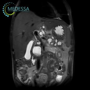
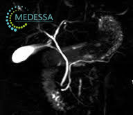
MRI of the Abdomen and Biliary Tract
- Unexplained abdominal pain or discomfort.
- Unintentional weight loss or appetite changes.
- Abnormal liver or gallbladder laboratory results.
- Suspected tumors, cysts, or other masses.
- Gallstone disease.
- Inflammatory conditions (cholecystitis, cholangitis).
- Preoperative assessment.
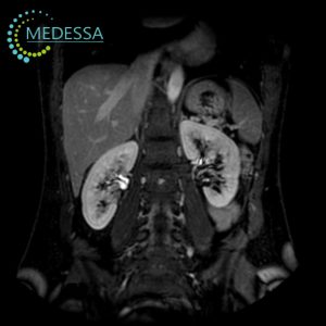
MRI of the Retroperitoneal Space
- Lower back or abdominal pain.
- Kidney function abnormalities.
- Suspected tumors or developmental abnormalities.
- Inflammatory diseases (pyelonephritis, hydronephrosis).
- Pre-surgical evaluation.
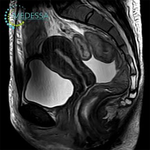
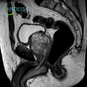
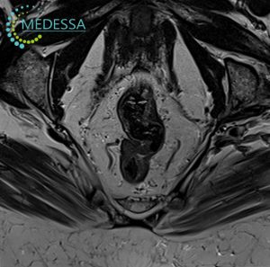
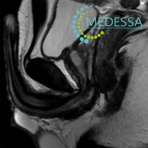
Pelvic MRI
For women:
- Lower abdominal or pelvic pain.
- Menstrual irregularities.
- Suspected endometriosis, fibroids, ovarian cysts.
- Infertility.
- Preoperative gynecological assessment.
For men:
- Urinary disorders.
- Pelvic or perineal pain.
- Suspected prostate diseases.
- Inflammatory urogenital conditions.
Rectal MRI
- Blood in stool or bowel habit changes.
- Anal or rectal discomfort.
- Suspected tumors or polyps.
- Cancer staging and assessment.
- Preoperative planning.
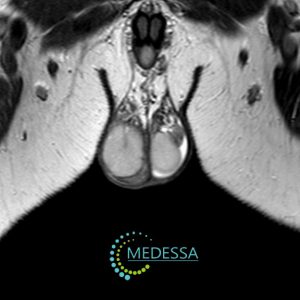
Scrotal MRI
- Pain or swelling.
- Suspected tumors or cysts.
- Inflammatory conditions (orchitis, epididymitis).
- Trauma evaluation.
Penile MRI
- Pain or deformity.
- Traumatic injury.
- Suspected masses.
- Peyronie’s disease or erectile dysfunction.
- Preoperative planning.
MRI with Contrast Enhancement
Contrast agents may be used for improved visualization of tumors, inflammatory changes, and vascular abnormalities. The decision to administer contrast is made individually by the physician.
Preparation for Internal Organ MRI
Dietary adjustments may be required before abdominal and pelvic MRI to reduce intestinal gas formation.
Please уточняйте preparation details when scheduling the examination.
Medical Documentation
Patients are advised to bring previous imaging results (ultrasound, CT, X-ray, DICOM files, medical reports) for comprehensive evaluation.
How the Procedure Is Performed
The patient lies on a movable table that slides into the MRI scanner. Remaining still during the scan is essential for image quality. The procedure is painless and monitored by medical staff.
Results
After the examination, patients receive printed images and digital DICOM files. The radiology report is provided within the established timeframe.
Contraindications
Absolute contraindications include pacemakers, certain metallic implants, neurostimulators, and other devices incompatible with MRI. The final decision is made by the physician.