
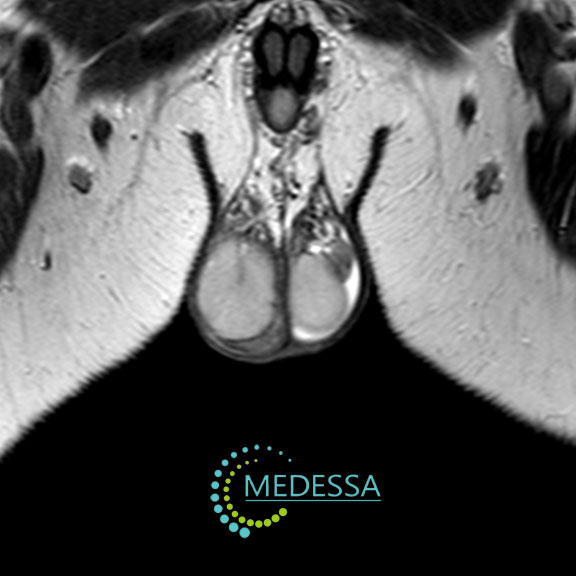
MRI of the Scrotum and Testicles
MRI of the scrotum and testicles is a non-invasive imaging technique that provides detailed visualization of the scrotal organs using magnetic fields and radiofrequency waves.
The examination allows evaluation of the testicles, epididymis, spermatic cord, and surrounding soft tissues.
What MRI Can Detect
- testicular tumors;
- cysts and masses;
- inflammatory processes;
- traumatic injuries;
- congenital abnormalities;
- vascular or perfusion disorders.
Indications
Pain or discomfort
MRI may be recommended to determine the cause of persistent pain or discomfort in the scrotal area.
Cryptorchidism
The method helps identify the location of an undescended testicle.
Suspected tumors
MRI can detect masses within the scrotum and determine their characteristics.
Trauma
Used to evaluate the extent of injury to scrotal tissues.
Inflammatory conditions
MRI can detect orchitis, epididymitis, and other inflammatory disorders.
Congenital anomalies
The examination helps identify structural abnormalities of the scrotum and testicles.
MRI of the scrotum is a valuable diagnostic tool that helps establish an accurate diagnosis and guide appropriate treatment.