
MRI of the Female Pelvic Organs
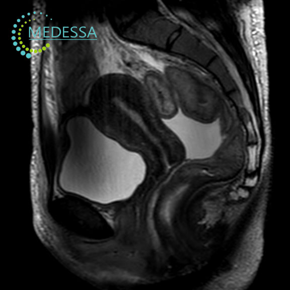
MRI of the female pelvic organs is a non-invasive imaging method used to obtain detailed visualization of the uterus, ovaries, fallopian tubes, bladder, and surrounding tissues.
The examination is widely used in gynecology to detect inflammatory conditions, cysts, tumors, and other pelvic abnormalities.
Indications for Pelvic MRI
Unclear results from other examinations
MRI may be recommended to clarify ultrasound findings or other diagnostic results.
Pelvic pain or discomfort
The study helps determine the cause of pain or discomfort in the pelvic region.
Suspected pelvic masses
MRI allows evaluation of pelvic masses, including their structure, size, and location.
Post-treatment follow-up
The examination is used to monitor treatment results and assess disease progression.
Conditions detected by pelvic MRI
Uterine fibroids
MRI accurately evaluates the size and location of fibroids within the uterus.
Endometriosis
The method helps detect endometriotic lesions and determine disease extent.
Ovarian cysts
MRI can differentiate between different types of ovarian cysts.
Ovarian tumors
The examination is used to identify benign and malignant ovarian tumors.
Bladder tumors
MRI helps detect masses in the urinary bladder and evaluate their spread.
MRI of the female pelvis is an important diagnostic tool that helps physicians establish an accurate diagnosis and plan appropriate treatment.