
MRI of Internal Organs in Chornomorsk (1.5 Tesla)
MRI of internal organs is a modern non-invasive imaging method used to assess the abdominal organs, retroperitoneal space, pelvic organs, and selected soft-tissue structures without ionizing radiation. MRI is widely used to detect tumors, inflammatory changes, cysts, structural abnormalities, traumatic consequences, biliary and urinary outflow disorders, and other clinically important abnormalities.
Chornomorsk
Phones:
Messengers:
Address: Chornomorsk, 6 Oleksandriyska Street
Working hours: 8:00 AM – 8:00 PM
At MEDESSA in Chornomorsk, internal organ MRI is performed on a 1.5 Tesla scanner. This field strength is widely regarded as the standard for routine MRI examinations and provides high-quality visualization of the liver, gallbladder, bile ducts, pancreas, spleen, kidneys, adrenal glands, pelvic organs, rectum, scrotum, and surrounding soft tissues.
What MRI of Internal Organs Can Show
MRI helps detect a broad range of common and less common conditions, including:
- benign and malignant tumors;
- metastatic lesions;
- cysts and other focal masses;
- inflammatory changes;
- congenital and acquired structural abnormalities;
- postoperative changes;
- biliary or urinary tract obstruction;
- lymph node and surrounding soft-tissue abnormalities.
When MRI of Internal Organs Is Recommended
MRI may be recommended in patients with pain, discomfort, unexplained laboratory abnormalities, suspected tumors, cysts, inflammatory disease, or when ultrasound and CT findings require further clarification.
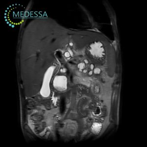
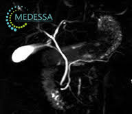
MRI of the Abdomen and Biliary Tract
This examination provides detailed assessment of the liver, gallbladder, bile ducts, pancreas, spleen, and adjacent structures. It is commonly used when a more accurate evaluation of masses, inflammatory disease, and biliary obstruction is needed.
- unexplained abdominal pain or discomfort;
- unintentional weight loss;
- appetite changes;
- abnormal liver or biliary laboratory results;
- suspected tumors, cysts, or other masses;
- gallstone-related disease and biliary obstruction;
- inflammatory conditions such as cholecystitis or cholangitis;
- preoperative or postoperative assessment.
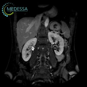
MRI of the Retroperitoneal Space
Retroperitoneal MRI helps evaluate the kidneys, adrenal glands, ureters, vascular and lymphatic structures, and surrounding tissues. It is useful for suspected inflammation, tumors, congenital anomalies, hydronephrosis, and other pathological changes.
- lower back or abdominal pain;
- kidney function abnormalities;
- suspected tumors or developmental abnormalities;
- inflammatory disease, including pyelonephritis;
- suspected hydronephrosis;
- pre-surgical clarification of anatomy and pathology.
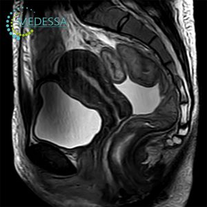
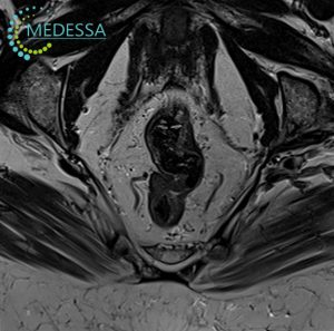
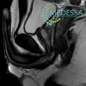
Pelvic MRI
Pelvic MRI is used for detailed assessment of reproductive, urinary, and surrounding soft-tissue structures in both women and men. It is especially valuable when precise anatomical definition is needed.
For women
- lower abdominal or pelvic pain;
- menstrual irregularities;
- suspected endometriosis;
- uterine fibroids;
- ovarian cysts;
- infertility workup;
- preoperative gynecologic assessment.
For men
- urinary symptoms;
- pelvic or perineal pain;
- suspected prostate disease;
- inflammatory urogenital conditions;
- pre-treatment assessment of disease extent.
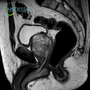
Rectal MRI
Rectal MRI is particularly important for local staging, evaluation of tumor spread, treatment planning, and preoperative assessment.
- blood in the stool;
- change in bowel habits;
- anal or rectal discomfort;
- suspected tumors or polyps;
- cancer staging and local extent assessment;
- preoperative planning.
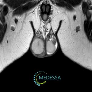
Scrotal MRI
Scrotal MRI may be used as a problem-solving examination when a more detailed evaluation of the testes, epididymis, coverings, and surrounding tissues is required.
- pain or swelling;
- suspected tumors or cysts;
- inflammatory conditions such as orchitis or epididymitis;
- assessment after trauma.
Penile MRI
This examination may help clarify traumatic, inflammatory, or structural abnormalities and can be useful before surgery.
- pain or deformity;
- traumatic injury;
- suspected masses;
- Peyronie’s disease;
- preoperative planning.
MRI with Contrast Enhancement
Contrast-enhanced MRI may be recommended to improve visualization of tumors, inflammatory changes, vascular abnormalities, and postoperative findings. MRI commonly uses gadolinium-based contrast agents, and the decision to administer contrast is made individually by the physician based on the clinical question and patient safety factors.
Why 1.5 Tesla MRI Is a Good Choice
1.5 Tesla MRI offers high diagnostic accuracy and detailed soft-tissue imaging for abdominal, pelvic, and retroperitoneal examinations. This field strength is widely used in routine clinical MRI practice for standard internal organ studies.
Preparation for Internal Organ MRI
Preparation depends on the body area being examined. Many MRI studies require no special preparation, but abdominal and pelvic MRI often involve temporary fasting and may require dietary adjustments to reduce bowel gas. Exact preparation instructions should be confirmed when scheduling the examination.
Medical Documentation
Patients are advised to bring previous imaging results, including ultrasound, CT, X-ray reports, DICOM files, and medical documentation. Comparing prior studies can improve the quality and clinical value of the final interpretation.
How the Procedure Is Performed
The patient lies on a movable table that slides into the MRI scanner. Remaining still during image acquisition is essential for high-quality images. The examination is painless and monitored by medical staff throughout the procedure.
Results
After the examination, patients receive images and a digital DICOM archive. The radiology report is issued within the center’s standard reporting timeframe and may vary depending on exam complexity.
Contraindications
Absolute or major contraindications may include pacemakers, certain metallic implants, neurostimulators, and other MRI-incompatible devices. The final decision about MRI safety is made by the physician after reviewing the safety questionnaire and the patient’s medical history.