
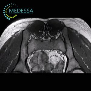
Spine MRI
Magnetic Resonance Imaging (MRI) is the gold standard for diagnosing spine conditions due to its non-invasive and highly informative nature. This method is completely safe and does not use radiation, offering an advantage over X-rays and CT scans. MRI allows detailed evaluation of intervertebral discs, ligaments, muscles, nerve roots, blood vessels, spinal cord membranes, and other structures.
MRI helps detect the following conditions:
- Benign and malignant spine tumors;
- Metastases of malignant tumors, their size and precise location;
- Multiple sclerosis;
- Inflammatory processes in the spinal cord;
- Osteochondrosis;
- Protrusions and herniated discs;
- Spinal joint deformities;
- Congenital and acquired spinal deformities.
Odesa
Phones:
Messengers:
Address: Odesa, 10 Kosmonavtiv Street
Working hours: 8:00 AM – 10:00 PM
Chornomorsk
Phones:
Messengers:
Address: Chornomorsk, 6 Oleksandriyska Street
Working hours: 8:00 AM – 8:00 PM
Indications for spine MRI:
- Cracking sounds with discomfort during bending and straightening;
- Acute or chronic back pain;
- Reduced sensation or numbness in upper or lower limbs.
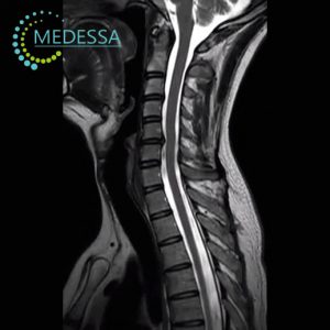
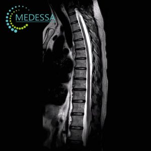
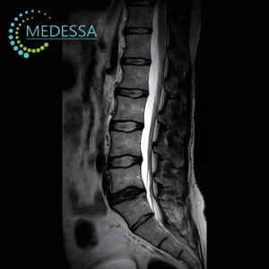
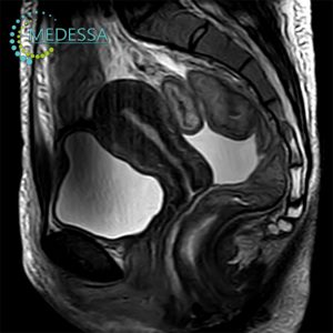
MRI of different spinal regions:
Cervical spine:
- Headache, dizziness;
- Fatigue, drowsiness, or insomnia;
- Numbness in upper limbs;
- Tinnitus;
- Stiffness and limited head mobility.
Thoracic spine:
- Numbness in upper limbs and fingers;
- Tightness in the chest;
- Muscle spasms;
- Pain and burning in the shoulder blades;
- Pain along intercostal nerves;
- Limited mobility of the thoracic spine;
- Spinal deformities.
Lumbar spine:
- Acute or chronic lower back pain radiating to the leg;
- Morning stiffness;
- Reduced sensation and numbness in lower limbs;
- Loss of sensation in the perineal area.
Sacral-coccygeal spine:
- Pain or discomfort in the sacrum and coccyx;
- Numbness or tingling;
- Suspected injuries or pathologies (fractures, tumors, inflammation);
- Pain radiating to other body parts;
- Deformities and changes in bowel or bladder function.
Contrast-enhanced MRI:
Contrast MRI is performed on a doctor’s recommendation when standard scans do not provide enough information. Safe gadolinium-based agents are injected intravenously to improve visualization of tumors and pathologies.
Preparation for MRI:
No special preparation is required. Avoid creams, gels, deodorants on the scan area. For contrast MRI, fasting is recommended.
- Filling out a questionnaire about possible contraindications;
- Providing results of previous examinations (MRI, CT, ultrasound);
- Removing all metal objects in the changing room.
How MRI is performed:
The patient is positioned on a movable table that slides into the scanner tunnel. Absolute stillness is required. Earplugs or headphones are provided for comfort. A qualified technician monitors the procedure via intercom.
MRI results:
Selected images are provided on paper, full electronic archive in DICOM format. MRI interpretation is available within 24 hours or later in complex cases. At MEDESSA, two experienced radiologists analyze scans for maximum accuracy.
Contraindications:
MRI is not performed in the presence of metallic implants or devices (pacemaker, neurostimulator, clips, ferromagnetic prostheses). Final decision is made by the MRI physician.