
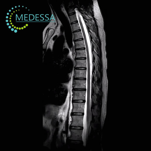
MRI of the Thoracic Spine
MRI of the thoracic spine is an advanced diagnostic imaging method used to obtain detailed images of the thoracic vertebrae, intervertebral discs, spinal cord, and surrounding soft tissues.
The examination uses magnetic fields and radio waves to produce high-resolution images without ionizing radiation. MRI is widely used to diagnose spinal injuries, degenerative disorders, inflammation, and tumors.
Structures evaluated during MRI
Vertebrae
The condition, structure, and alignment of the thoracic vertebrae are assessed.
Intervertebral discs
MRI helps detect disc protrusion, disc herniation, and degenerative disc disease.
Ligaments and muscles
Soft tissues, ligaments, and muscles surrounding the thoracic spine are evaluated.
Spinal cord and nerve roots
The examination evaluates the spinal cord and nerve roots to detect compression or inflammation.
Conditions detected by MRI
- intervertebral disc herniation;
- disc protrusion;
- degenerative spine disease;
- spinal canal stenosis;
- inflammatory disorders;
- spinal tumors;
- post-traumatic changes.
Indications for MRI
- pain in the thoracic spine;
- pain between the shoulder blades;
- numbness or weakness in the limbs;
- limited spinal mobility;
- suspected disc herniation;
- spinal injuries;
- suspected tumors or inflammatory diseases.
MRI of the thoracic spine provides detailed visualization of spinal structures and helps physicians establish an accurate diagnosis and treatment plan.