
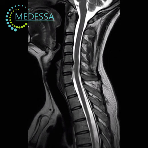
MRI of the Cervical Spine
MRI of the cervical spine is a modern diagnostic imaging technique used to obtain detailed images of the cervical vertebrae, intervertebral discs, nerve roots, and surrounding soft tissues.
The examination uses magnetic fields and radio waves to produce high-resolution images without ionizing radiation. MRI is one of the most effective methods for diagnosing spinal disorders.
Structures evaluated during MRI
Vertebrae, muscles and ligaments
The condition of the cervical vertebrae, ligaments, and muscles is assessed to detect injury, deformity, or inflammation.
Intervertebral discs
MRI helps detect disc protrusion, disc herniation, and other degenerative disc changes.
Spinal canal
The examination evaluates the spinal canal and possible compression of neural structures.
Nerve roots and spinal cord
MRI allows detailed evaluation of the spinal cord and nerve roots.
Conditions detected by MRI
- intervertebral disc herniation;
- disc protrusion;
- degenerative spine disease;
- spinal canal stenosis;
- inflammatory processes;
- spinal tumors;
- post-traumatic changes.
Indications for MRI
- neck pain;
- headache or dizziness;
- numbness or weakness in the arms;
- limited neck mobility;
- suspected disc herniation;
- spinal injuries;
- suspected tumors or inflammatory diseases.
MRI of the cervical spine provides detailed visualization of spinal structures and helps physicians determine an accurate diagnosis and treatment plan.