
Doctors of Medessa Medical Center in Odesa
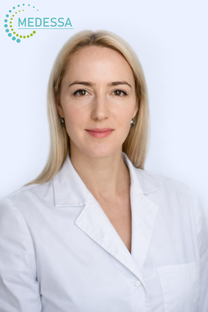
Nataliia Grubnyk
Ophthalmologist of the highest qualification category, PhD in Medical Sciences
- Graduated from Bogomolets National Medical University (2007)
- Specialization: cataract and vitreoretinal surgery, reconstructive eye trauma surgery
- Member of the Ukrainian Society of Vitreoretinal Surgeons
- Member of the Ukrainian Society of Ophthalmologists
- Member of the European Society of Cataract and Refractive Surgeons (ESCRS)
- Member of the European Society of Retina Specialists (EURETINA)
- 2007–2023 — Ophthalmic surgeon at the Filatov Institute of Eye Diseases
- Since 2023 — Vitreoretinal and cataract surgeon at Mykolaiv Regional Hospital
Eye diagnostics with modern equipment
Comprehensive eye examination using high-precision equipment for accurate diagnosis
Examinations using modern equipment at the Medessa medical center allow our ophthalmologists to obtain precise information about each patient’s eye condition.
Every case is unique and requires an individual approach to choosing the appropriate treatment.
Comprehensive eye diagnostics allows to:
- Assess the condition of the eyes and visual functions
- Make an accurate diagnosis
- Prescribe a treatment program
- Determine the need for surgical intervention
When is an eye examination necessary?
- Noticed sudden or gradual vision loss, or missing parts of the visual field.
- All patients with myopia, astigmatism, hyperopia, glaucoma, cataract, retinal or optic nerve disorders, etc.
- All adults — once a year.
- Experience discomfort in the eyes, dryness, redness, tearing, or “sand” sensation.
- Have diabetes, hypertension, or other chronic conditions.
- Your relatives suffer from glaucoma.
- During pregnancy.
- After 40 years, even if there are no vision complaints.
- Regularly use glasses or contact lenses.
- Planning eye surgery.
Medessa Medical Center offers the following diagnostics
- Visometry (assessment of visual acuity and its characteristics)
- Autorefractokeratometry
- Pneumotonometry
- Icare tonometry
- Biomicroscopy
- Ophthalmoscopy — examination of the fundus using high-diopter lenses (90 D)
- Gonioscopy with Goldman lens
- Computer perimetry
- Optical coherence tomography (OCT) of the macula and optic nerve
Medessa Medical Center conducts ophthalmological examinations using modern equipment from leading global brands.
Stages of a Comprehensive Eye Examination
- Assessment of refraction and visual acuity (autorefractokeratometry and visometry).
- Measurement of intraocular pressure (pneumotonometry, Icare tonometry).
- Biomicroscopy of the eyelids, anterior segment, and optical media.
- Pupil dilation.
- Ophthalmoscopy (examination of the fundus, retina, and optic disc).
- Consultation with an ophthalmologist, diagnosis, and treatment recommendations.
- Consultative report including all examination data, treatment plan, and/or follow-up instructions.
If further clarification of the diagnosis is needed, additional tests such as computer perimetry and optical coherence tomography (OCT) may be recommended.
How the Primary Comprehensive Eye Examination is Conducted
- You arrive at the Medessa medical center at the scheduled time.
- You are invited to a completely contactless and painless eye examination, followed by intraocular pressure measurement.
- Next, eye drops are instilled to dilate the pupils (medicated mydriasis) so the ophthalmologist can examine the retina and optic disc. Vision may be blurry for the rest of the day. Driving is not recommended; sunglasses are advised.
- You sit for 10–20 minutes with your eyes closed while the pupils dilate.
- The doctor takes your medical history and examines the anterior and posterior segments of the eye using a slit lamp.
- The ophthalmologist provides a report with recommendations and a treatment plan or refers you for additional examinations if necessary.
Особливості підготовки пацієнта
- Після обстеження ваш зір зблизька і вдалину буде трохи нечітким, тому читати й працювати за комп’ютером буде складно ще декілька годин.
- Сідати за кермо не рекомендуємо.
- Радимо взяти з собою сонцезахисні окуляри, щоб після обстеження не відчувати дискомфорту від яскравого сонячного світла.
Diagnostic Methods
- Visometry — assessment of central visual acuity.
- Autokeratorefractometry — method to determine the eye’s refraction and corneal refractive power.
- Biomicroscopy — examination of the anterior and posterior segments of the eye using a slit lamp.
- Ophthalmoscopy — examination of the retina and optic disc.
- Gonioscopy — examination of the anterior chamber angle.
- Pneumotonometry, Icare tonometry — measurement of intraocular pressure. Recommended for all adults over 40 years.
- Computer perimetry — functional method to assess the optic nerve and retina. Detects visual field defects caused by glaucoma, retinal disease, or neuro-ophthalmological conditions.
- Optical coherence tomography (OCT) — painless, fast, non-invasive examination of the retina and optic disc. Allows evaluation of morphological and morphometric features. Requires no special preparation or pupil dilation. Used for precise diagnosis of retinal and optic nerve diseases.
| Service Name | Cost (UAH) |
|---|---|
| Promotional Package: Diagnostic Package 1 – basic vision diagnostics + ophthalmologist consultation (visometry, autorefractometry, tonometry, ophthalmoscopy, biomicroscopy) | 1000 |
| Promotional Package: Diagnostic Package 2 – Diagnostic Package 1 + OCT of both eyes | 1500 |
| Promotional Package: Diagnostic Package 3 – Diagnostic Package 2 + perimetry of both eyes | 2200 |
| Ophthalmologist consultation (urgent care for conjunctivitis, foreign bodies, eye injuries) without diagnostics | 1000 |
| Ophthalmologist consultation (“second opinion”, without diagnostics) | 700 |
| OCT (Optical Coherence Tomography of the macula and optic nerve head) – both eyes | 1000 |
| OCT (Optical Coherence Tomography of the macula and optic nerve head) – one eye | 600 |
| Eyeglasses prescription (autorefractometry) | 400 |
| Tonometry (pneumotonometry, Icare tonometry) | 400 |
| Computerized perimetry – both eyes | 800 |
| Computerized perimetry – one eye | 450 |
| Follow-up monitoring of glaucoma patients (eye drop adjustment) | 600 |
| Schirmer test (for dry eye syndrome diagnosis) | 200 |
| Pachymetry – both eyes | 300 |