
MRI of Internal Organs in Odesa 3.0 Tesla — Accurate and Safe Diagnostic Imaging
MRI of internal organs is a modern non-invasive imaging method that provides highly detailed assessment of the abdominal organs, retroperitoneal space, pelvic organs, and surrounding soft tissues without ionizing radiation. This examination helps detect both common and complex abnormalities at an early stage, characterize tumors and cystic lesions, assess inflammatory changes, and support treatment planning.
Odesa
Phones:
Messengers:
Address: Odesa, 10 Kosmonavtiv Street
Working hours: 8:00 AM – 10:00 PM
At MEDESSA in Odesa, examinations are performed on a high-field 3.0 Tesla MRI system — one of the strongest MRI scanners in Ukraine, among the first systems of this class in the country and the first on southern Ukraine. The combination of high magnetic field strength and optimized imaging protocols provides excellent soft-tissue contrast and outstanding diagnostic value for liver imaging, biliary assessment, pelvic MRI, tumor evaluation, and complex multi-organ cases.
What MRI of internal organs can evaluate
MRI allows detailed visualization of:
- the liver;
- the gallbladder;
- the bile ducts;
- the pancreas;
- the spleen;
- the kidneys and adrenal glands;
- the ureters and adjacent tissues;
- the female pelvic organs;
- the prostate, seminal vesicles, and adjacent male pelvic structures;
- the rectum and surrounding mesorectal tissues;
- the scrotum and perineal soft tissues in selected cases;
- vascular and lymphatic structures within the examined region.
When MRI of internal organs is recommended
MRI is especially useful when highly detailed soft-tissue assessment is required, when ultrasound or CT findings need clarification, or when precise local staging is important for treatment decisions. It is commonly used in the diagnostic workup of tumors, inflammatory disease, congenital abnormalities, complex cystic lesions, postoperative changes, and preoperative planning.
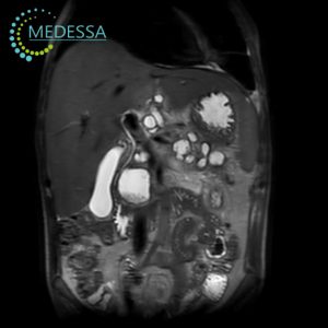
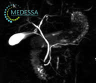
Abdominal MRI and biliary system MRI
Abdominal MRI provides detailed evaluation of the liver, gallbladder, bile ducts, pancreas, spleen, abdominal soft tissues, and regional lymph nodes. In selected cases, MR cholangiopancreatography (MRCP) can be added as a dedicated non-invasive MRI technique for detailed visualization of the biliary and pancreatic ductal systems.
Indications for abdominal and biliary MRI
- unexplained abdominal pain or persistent abdominal discomfort;
- unintentional weight loss or appetite changes;
- abnormal liver function tests;
- suspected liver tumors, cysts, or focal lesions;
- assessment of the gallbladder and bile ducts;
- suspected inflammatory conditions such as cholecystitis or cholangitis;
- evaluation of pancreatic abnormalities;
- preoperative planning and follow-up after treatment.
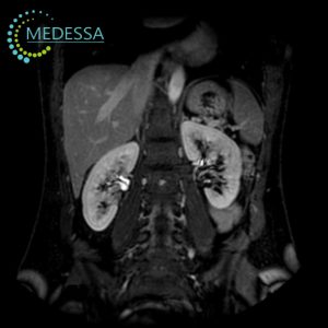
MRI of the retroperitoneal space
Retroperitoneal MRI is used to assess the kidneys, adrenal glands, ureters, vessels, lymph nodes, and surrounding retroperitoneal soft tissues. It is valuable for characterizing masses, inflammatory disease, congenital anomalies, and post-traumatic or postoperative changes.
Indications for retroperitoneal MRI
- flank pain, lower back pain, or unexplained abdominal symptoms;
- suspected renal, adrenal, or retroperitoneal tumors;
- developmental anomalies of the urinary tract;
- complex inflammatory disorders;
- suspected urinary obstruction or collecting system dilatation;
- follow-up after treatment;
- pre-surgical evaluation.
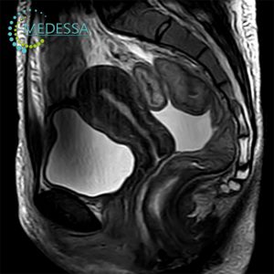
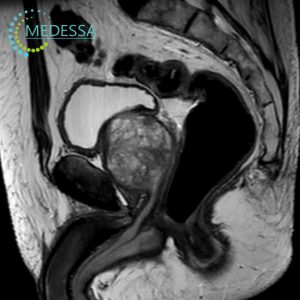
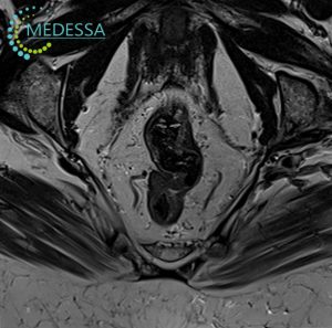
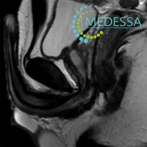
Pelvic MRI
Pelvic MRI is one of the most informative imaging methods for assessment of the uterus, ovaries, adnexal structures, bladder, rectum, prostate, seminal vesicles, and surrounding pelvic soft tissues. High-resolution 3.0 Tesla MRI is particularly useful for evaluating small lesions, infiltrative disease, and local tumor spread.
Pelvic MRI for women
- pelvic pain or lower abdominal pain;
- menstrual irregularities;
- suspected endometriosis;
- suspected uterine fibroids, adenomyosis, ovarian cysts, or other adnexal masses;
- infertility as part of a broader diagnostic workup;
- clarification of abnormal ultrasound or CT findings;
- preoperative gynecologic assessment;
- follow-up after treatment or surgery.
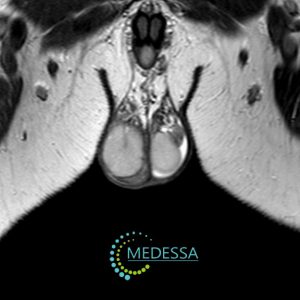
Pelvic MRI for men
- urinary symptoms;
- pelvic, suprapubic, or perineal pain;
- suspected prostate disease;
- assessment of the prostate and seminal vesicles;
- inflammatory disorders of the urogenital system;
- local staging before treatment;
- post-treatment follow-up.
Rectal MRI
Rectal MRI is a key method for local staging of rectal cancer, allowing evaluation of tumor depth, mesorectal fascia, mesorectal fat, and regional lymph nodes. It also plays an important role in treatment planning, including surgery and radiation therapy, and may be useful in selected inflammatory or postoperative cases.
Indications for rectal MRI
- blood in the stool or persistent change in bowel habits;
- rectal pain, pressure, or discomfort;
- suspected rectal mass, tumor, or infiltrative lesion;
- local staging of rectal cancer;
- preoperative planning;
- follow-up after therapy.
Scrotal MRI
Scrotal MRI is typically a problem-solving examination rather than a first-line test. It may be recommended when ultrasound findings are inconclusive or when additional tissue characterization is required. MRI can help define the extent of a lesion, assess soft tissues, and clarify complex abnormalities.
Indications for scrotal MRI
- pain or scrotal enlargement of unclear origin;
- suspected tumor;
- characterization of cystic or solid lesions;
- assessment after trauma;
- complex inflammatory conditions;
- inconclusive ultrasound findings.
Penile MRI
Penile MRI is performed in selected cases when highly detailed anatomical assessment is necessary. It may be useful in traumatic injury, fibrotic plaque assessment, suspected masses, and preoperative planning in complex urologic conditions.
Indications for penile MRI
- pain or deformity;
- traumatic injury;
- suspected mass lesion;
- evaluation of Peyronie’s disease in selected cases;
- preoperative planning.
Contrast-enhanced MRI
Contrast enhancement may be used when additional lesion characterization is needed, especially for tumors, inflammatory processes, vascularized lesions, postoperative assessment, and clarification of equivocal findings. Gadolinium-based contrast agents are administered only when clinically indicated and after individual safety assessment, including consideration of renal function when appropriate.
Preparation for MRI of internal organs
Preparation depends on the specific anatomical region being examined. For abdominal or pelvic MRI, patients may receive dietary instructions, recommendations to reduce bowel gas, or specific bladder filling guidance. Exact preparation instructions should always be clarified when scheduling the examination.
Medical documentation to bring
Previous medical records may significantly improve interpretation quality. Patients are encouraged to bring:
- prior ultrasound, CT, MRI, or X-ray reports;
- DICOM discs or electronic image archives;
- hospital discharge summaries;
- relevant laboratory results;
- referral notes and previous specialist consultations.
How the procedure is performed
The patient lies on a movable table that slides into the MRI scanner. Remaining still is essential for image quality, and some protocols may require short breath-holds. The examination is painless and continuously monitored by the medical staff through an intercom system.
MRI results
After the examination, patients receive image data and a DICOM archive together with a written radiology report. In complex cases, additional expert review may be used to ensure the most accurate and clinically useful interpretation.
Contraindications
Absolute and relative contraindications are assessed individually. Important limitations may include certain pacemakers, some neurostimulators, specific metallic implants, vascular clips, and other devices that are not compatible with MRI. The final decision about whether MRI can be safely performed is made by the physician after reviewing the MRI safety questionnaire and available medical documentation.