
Spine MRI in Odesa 3.0 Tesla
Spine MRI is an advanced diagnostic imaging method that allows detailed visualization of vertebrae, intervertebral discs, spinal cord, nerve roots, ligaments, muscles and surrounding soft tissues. MRI does not use ionizing radiation and is considered one of the safest diagnostic tools in modern medicine.
Odesa
Phones:
Messengers:
Address: Odesa, 10 Kosmonavtiv Street
Working hours: 8:00 AM – 10:00 PM
Advantages of 3.0 Tesla MRI
High-field 3.0 Tesla MRI provides superior image resolution compared to lower field systems. This allows early detection of structural abnormalities, inflammatory diseases, tumors and degenerative spine disorders. MEDESSA in Odesa operates one of the first 3 Tesla MRI systems in Ukraine and the first high-field system of this level in southern Ukraine.
- high-definition visualization of intervertebral discs;
- detailed assessment of spinal cord structures;
- early detection of herniated discs;
- identification of inflammatory diseases;
- accurate tumor diagnostics;
- optimized MRI protocols.
Structures visualized during spine MRI
- vertebral bodies;
- intervertebral discs;
- spinal cord;
- nerve roots;
- ligaments;
- facet joints;
- muscles;
- blood vessels;
- meninges.
Conditions detected by spine MRI
- degenerative disc disease;
- disc herniation;
- disc protrusion;
- spinal stenosis;
- multiple sclerosis;
- tumors;
- metastases;
- traumatic injuries;
- inflammatory diseases;
- nerve compression;
- congenital abnormalities.
Indications for spine MRI
- back pain;
- neck pain;
- radiating pain to arms or legs;
- muscle weakness;
- numbness or tingling;
- limited mobility;
- post-traumatic evaluation.
MRI of different spine regions
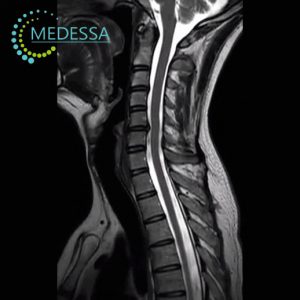
Cervical spine
- headache;
- dizziness;
- neck stiffness;
- upper limb numbness.
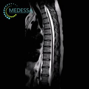
Thoracic spine
- pain between shoulder blades;
- postural abnormalities;
- intercostal neuralgia.
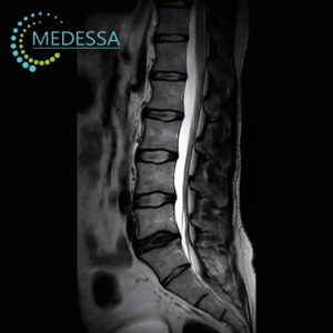
Lumbar spine
- lower back pain;
- sciatica;
- leg weakness.
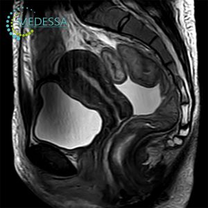
Sacral spine
- sacral pain;
- coccyx trauma;
- neurological symptoms.
Contrast-enhanced MRI
Gadolinium contrast agents improve visualization of tumors, inflammatory lesions and vascular abnormalities. Contrast MRI increases diagnostic confidence.
Preparation
- remove metallic objects;
- inform physician about implants;
- fasting recommended before contrast MRI.
Procedure
The patient lies still during scanning while the technologist monitors the examination.
Results
DICOM images and radiology report are provided after examination.
Contraindications
Pacemakers, ferromagnetic implants and certain electronic devices.