


MRI 3.0 Tesla
THE FIRST IN ODESSA
AND IN SOUTHERN UKRAINE
The center is equipped with a modern
magnetic resonance
tomograph Philips Achieva with
a magnetic field of 3.0 Tesla

TEAM OF EXPERIENCED
PROFESSIONALS
We are proud to have
a united team of
highly qualified
like-minded doctors
and attentive staff

CONVENIENCE AND
COMFORT
Comfortable and sunny
rooms with short waiting times.
We are here to make it easy for
you at every stage of
MRI diagnostics

AFFORDABLE
PRICES
MRI 3.0
Affordable prices,
matching the level and quality
of examinations
MRI in Odessa
Our Doctors
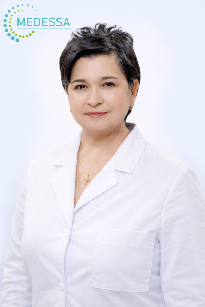
Bondar Eleonora Leonidivna
Radiologist of the Highest Qualification Category
Professional Experience
- Over 35 years of professional experience
- Specialist in MRI and CT diagnostics
- Member of the Ukrainian and European Radiological Associations
- Primary focus — oncology diagnostics and monitoring

Kovalyov Borys Oleksandrovych
Radiologist of the Highest Qualification Category
Professional Experience
- Over 45 years of professional experience
- Specialist in MRI and CT diagnostics
- Member of the Ukrainian and European Radiological Associations
- One of the first MRI and CT diagnostic physicians in Odesa

Borozenko Kseniia Olehivna
Radiologist of the Highest Qualification Category
Professional Experience
- Over 20 years of professional experience
- Specialist in MRI and CT diagnostics
- Member of the Ukrainian and European Radiological Associations
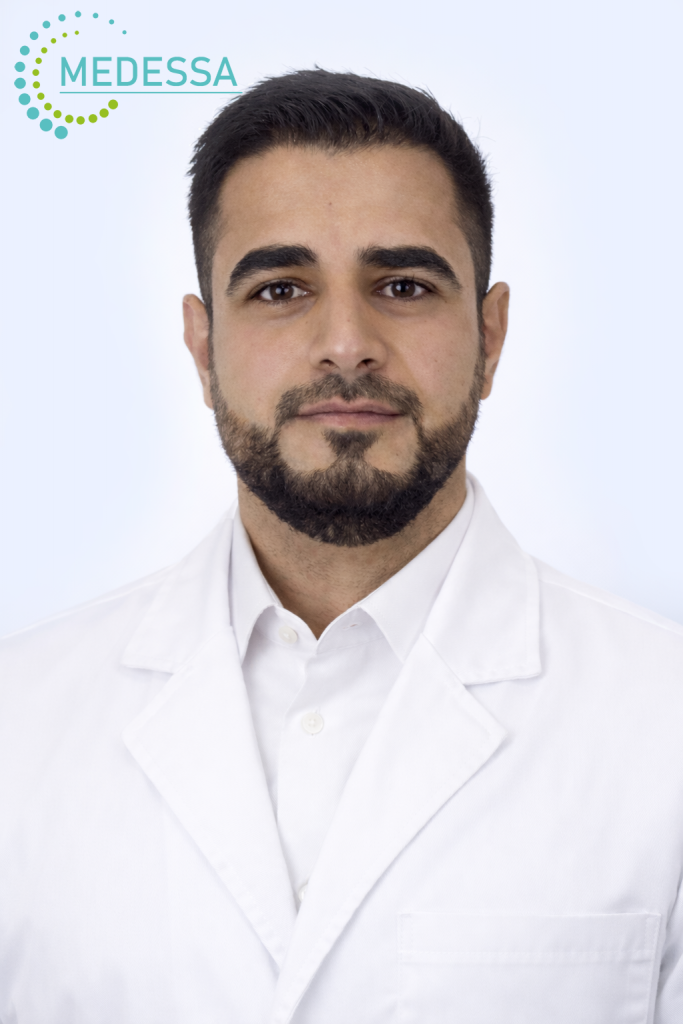
Al-Gburi Ahmed
Radiologist, Clinical Resident specializing in Radiology
Professional Experience
- Over 5 years of professional experience
- Specialist in MRI, CT, and X-ray diagnostics
- Member of the Ukrainian and European Radiological Associations
- Fluent in 4 languages

Hrubnyk Nataliia Pavlivna
Ophthalmologist of the Highest Qualification Category, PhD in Medical Sciences
Education
Graduated from Bogomolets National Medical University in 2007.
Specialization
- Cataract surgery
- Vitreoretinal surgery
- Reconstructive surgery for ocular trauma
Professional Memberships
- Member of the Ukrainian Society of Vitreoretinal Surgeons
- Member of the Ukrainian Society of Ophthalmologists
- Member of the European Society of Cataract and Refractive Surgeons (ESCRS)
- Member of the European Society of Retina Specialists (EURETINA)
Professional Experience
- 2007–2023 — Ophthalmic surgeon, Department of Post-Traumatic Eye Pathology, Filatov Institute of Eye Diseases and Tissue Therapy, National Academy of Medical Sciences of Ukraine
- Since 2023 — Vitreoretinal and Cataract Surgeon, Mykolaiv Regional Hospital

Zolotaryova Kseniia Olehivna
Cardiologist of the Highest Qualification Category, PhD in Medical Sciences
Professional Activity
- Specialist in ultrasound diagnostics (Echocardiography, ECHO-CG)
- Faculty member of the Department of Internal Medicine, Odesa National Medical University
- Specializes in diagnosis, treatment, and prevention of cardiovascular diseases
International Memberships
- Member of the Ukrainian and European Associations of Cardiology
- Member of the Heart Failure Association (HFA), European Society of Cardiology (ESC), ID 1351838 since 15.07.2023
- Member of the European Association of Cardiovascular Imaging (EACVI), European Society of Cardiology (ESC), ID 1351838 since 15.07.2023

Zaletskyi Volodymyr Volodymyrovych
Ultrasound Diagnostic Physician and Obstetrician-Gynecologist of the Highest Qualification Category, PhD in Medical Sciences
Professional Experience
- Over 28 years of professional experience
- Specializes in pelvic ultrasound, abdominal studies, prenatal and postnatal ultrasound
- Performs a wide range of ultrasound diagnostics
Professional Memberships
- Member of the Ukrainian and European Association of Ultrasound Diagnostic Specialists
About the Doctor
An experienced specialist whom patients trust for precise and attentive ultrasound diagnostics.
Medical Center MEDESSA



BOOK YOUR MRI SCAN ON THE MOST POWERFUL MRI SCANNER IN ODESSA


MRI in Odessa with 3.0 Tesla scanner — MEDESSA
About MEDESSA Diagnostic Center
MEDESSA Diagnostic Center provides high-precision MRI examinations in Odessa using an expert-class Philips Achieva 3.0 Tesla scanner. A 3 Tesla magnetic field allows obtaining extremely detailed medical images with high spatial resolution, improving diagnostic accuracy even in early or subtle pathological changes.
The MRI 3.0 Tesla system installed in Odessa is one of the most powerful MRI scanners in Ukraine. It is among the first 3 Tesla MRI systems introduced in the country and the first 3.0 Tesla MRI system in southern Ukraine. Examination protocols are optimized according to international radiology standards to ensure highly informative diagnostic results.
What is Magnetic Resonance Imaging (MRI)
MRI is an advanced non-invasive diagnostic imaging method that produces detailed cross-sectional images of organs, soft tissues, blood vessels, nervous system structures, and musculoskeletal components without the use of ionizing radiation.
MRI is widely used for diagnosing:
- tumors and oncological diseases
- inflammatory processes
- degenerative spine disorders
- ligament and tendon injuries
- vascular abnormalities
- traumatic injuries
- neurological disorders
- congenital anatomical abnormalities
How MRI works
MRI technology is based on the interaction between a strong magnetic field and radiofrequency pulses affecting hydrogen atoms within body tissues. The scanner processes signals and reconstructs highly detailed anatomical images in multiple planes.
3 Tesla MRI provides:
- superior image clarity
- visualization of small anatomical structures
- detection of early-stage diseases
- advanced imaging techniques availability
- high diagnostic confidence
Advantages of 3.0 Tesla MRI in Odessa
High-field 3 Tesla MRI is considered a modern gold standard in diagnostic imaging. Increased magnetic field strength provides improved image quality compared to 1.5 Tesla scanners, especially for brain imaging, vascular studies, joint evaluation, spine diagnostics, and soft tissue assessment.
Why patients choose MEDESSA
- one of the most powerful MRI scanners in Ukraine — 3.0 Tesla
- first 3 Tesla MRI in southern Ukraine
- internationally optimized scanning protocols
- high diagnostic accuracy
- experienced radiologists
- comfortable examination conditions
- early disease detection
Early and accurate MRI diagnosis helps physicians establish precise treatment strategies and monitor therapy effectiveness, improving overall patient outcomes.
3.0 Tesla MRI in Odessa — highest diagnostic accuracy
What is 3.0 Tesla MRI
3.0 Tesla MRI is a high-field magnetic resonance imaging technology that produces highly detailed images of organs, soft tissues, blood vessels, and nervous system structures. The stronger magnetic field allows significantly improved image quality compared to lower field MRI systems.
The MEDESSA diagnostic center in Odessa is equipped with an expert-class Philips Achieva 3.0T scanner. This MRI system is one of the most powerful MRI scanners in Ukraine, among the first 3 Tesla systems installed in the country and the first 3.0 Tesla MRI scanner in southern Ukraine. Imaging protocols are optimized according to international radiology standards to achieve maximum diagnostic accuracy.
Advantages of 3.0 Tesla MRI
High-resolution anatomical imaging
3 Tesla MRI provides superior spatial resolution, allowing detailed visualization of the brain, pituitary gland, joints, spine, soft tissues, ligaments, blood vessels and internal organs. This allows detection of subtle pathological changes that may not be visible on lower field MRI scanners.
Accurate early disease detection
High-field MRI improves the detection of tumors, inflammatory diseases, demyelinating disorders, degenerative spine diseases, ligament injuries, vascular abnormalities and other pathological conditions at early stages.
Advanced vascular imaging
MR angiography with 3 Tesla MRI enables highly detailed visualization of arteries and veins, helping detect aneurysms, stenosis, thrombosis and vascular malformations without ionizing radiation.
Advanced MRI techniques
- MR spectroscopy
- DTI tractography
- diffusion imaging
- perfusion imaging
- functional MRI
High diagnostic confidence
3 Tesla MRI provides a high level of diagnostic reliability, especially in complex clinical cases requiring precise evaluation.
Why choose 3.0 Tesla MRI at MEDESSA
- one of the most powerful MRI scanners in Ukraine
- first 3 Tesla MRI system in southern Ukraine
- advanced imaging protocols
- high diagnostic accuracy
- early disease detection
- excellent soft tissue visualization
3 Tesla MRI is considered a modern gold standard in diagnostic imaging, allowing physicians to establish accurate diagnoses and select the most effective treatment strategies.
3.0 Tesla MRI with contrast in Odessa — enhanced diagnostic accuracy
Contrast-enhanced MRI 3.0 Tesla
Contrast-enhanced MRI significantly improves visualization of anatomical structures and pathological changes. Gadolinium-based contrast agents increase image clarity and allow more accurate assessment of tissues, blood vessels and tumors.
At MEDESSA diagnostic center in Odessa, MRI examinations are performed using Philips Achieva 3.0 Tesla scanner — one of the most powerful MRI systems in Ukraine and the first 3 Tesla MRI system in southern Ukraine. High magnetic field strength improves sensitivity to contrast enhancement.
When contrast MRI is recommended
Contrast agents are administered intravenously and accumulate in tissues with increased blood supply or altered metabolism. This allows better differentiation between normal and pathological tissues.
Contrast MRI is commonly used for evaluation of:
- tumors and metastases
- brain and pituitary disorders
- vascular abnormalities
- spine diseases
- joint pathology
- pelvic organs
- liver and kidney diseases
Advantages of contrast-enhanced 3 Tesla MRI
Early disease detection
Contrast enhancement allows detection of small lesions, tumors, inflammatory processes and vascular abnormalities.
Improved vascular visualization
Contrast-enhanced MR angiography provides detailed visualization of arteries and veins.
Higher diagnostic confidence
Contrast MRI improves diagnostic accuracy and helps determine treatment strategy.
Safety of gadolinium contrast agents
Modern gadolinium-based contrast agents are generally well tolerated and have a high safety profile.
Why choose contrast MRI at MEDESSA
- 3 Tesla MRI — one of the most powerful systems in Ukraine
- first 3 Tesla MRI in southern Ukraine
- high diagnostic accuracy
- advanced imaging protocols
- early detection of diseases
Contrast-enhanced MRI improves diagnostic precision and helps physicians make accurate clinical decisions.
Preparation for 3.0 Tesla MRI in Odessa
Proper preparation for a 3.0 Tesla MRI helps achieve the highest image quality, reduces the risk of artifacts, and makes the examination more comfortable for the patient. At MEDESSA diagnostic center in Odessa, MRI studies are performed using a high-field 3.0 Tesla scanner — one of the most powerful MRI systems in Ukraine — providing excellent visualization of anatomical structures, blood vessels, nervous tissue, joints, soft tissues, and internal organs.
3.0 Tesla MRI in Odessa is especially valuable when maximum diagnostic accuracy is required, including early detection of disease, characterization of tumors, and detailed assessment of the brain, spine, blood vessels, joints, soft tissues, abdominal organs, and pelvic structures. To obtain the most informative results, patients should follow the recommended preparation guidelines.
What to do before your appointment
It is recommended to arrive in advance to complete the necessary paperwork, fill out the MRI safety questionnaire, and inform the medical staff about any important medical details. Before MRI, you should tell the radiology team if you have:
- a pacemaker, neurostimulator, cochlear implant, or any electronic implanted device
- metal implants, clips, stents, vascular devices, or joint prostheses
- metal fragments in the body or near the eyes
- pregnancy or possible pregnancy
- claustrophobia, anxiety, or difficulty staying in enclosed spaces
- kidney disease or a history of allergic reactions if contrast-enhanced MRI is planned
- pain that may make it difficult to remain still during the scan
Clothing and personal items before MRI
You should come in comfortable clothing without metal components. Before entering the MRI room, all metallic objects and electronic devices must be removed because they may affect safety and image quality. This includes:
- jewelry, chains, earrings, and piercings
- watches, fitness trackers, smartphones, and bank cards
- glasses, hearing aids, and removable dental prostheses
- hairpins, metallic hair accessories, belts, and undergarments with metal parts
- clothing with zippers, buttons, hooks, or metallic inserts
If you are wearing patches, tapes, braces, orthopedic supports, or warming systems, you should inform the staff in advance. Some materials may contain metal or may heat up during MRI.
Medical documents to bring with you
For a more accurate interpretation of your MRI results, it is helpful to bring previous medical records if available:
- prior MRI, CT, ultrasound, or X-ray reports
- hospital discharge summaries
- specialist consultation notes
- referral for the examination
- information about previous surgeries or implanted devices
Comparison with prior imaging studies helps evaluate disease progression, treatment response, and newly developed changes.
Remaining still during the examination
Remaining completely still during MRI is essential. Even minor motion can reduce image sharpness and lower the diagnostic value of the examination. This is especially important for MRI of the brain, orbits, pituitary gland, spine, joints, blood vessels, and other small anatomical structures.
If you have pain, tremor, severe anxiety, or claustrophobia, this should be discussed in advance. In selected cases, additional preparation or sedative support may be considered when medically appropriate.
Preparation for MRI of the head, brain, and neck
Before MRI of the head, brain, orbits, pituitary gland, paranasal sinuses, or soft tissues of the neck, it is advisable not to use decorative cosmetics, especially products containing metallic particles, glitter, or long-lasting pigments. False eyelashes, metallic hair clips, and decorative hair accessories should also be removed.
These precautions help reduce artifacts and improve visualization quality in the scanned area.
Preparation for MRI of joints, limbs, and soft tissues
Before MRI of joints, ligaments, tendons, muscles, and soft tissues, it is advisable to remove braces, compression wraps, orthoses, kinesiology tape, warming patches, and other external support materials unless otherwise approved by your physician. Creams, ointments, and gels should not be applied directly to the area immediately before the scan.
For MRI of the knee, shoulder, hip, ankle, elbow, or wrist, the examined area should be as free as possible from foreign materials that may degrade image quality.
Preparation for abdominal, pelvic, and retroperitoneal MRI
Preparation for MRI of the abdomen, pelvis, retroperitoneal space, biliary system, or other specialized examinations may vary depending on the body area and the clinical purpose of the study. In many cases, patients may be advised to:
- avoid food for several hours before the examination if specifically instructed for that type of MRI
- temporarily limit foods that increase bowel gas when abdominal organs are being examined
- follow individualized instructions regarding bladder filling or emptying for pelvic MRI
The exact preparation plan depends on the specific type of MRI being performed. For this reason, patients should always follow the detailed instructions given for their particular examination.
Preparation for contrast-enhanced MRI
If contrast-enhanced MRI is planned, patients should report kidney disease, previous allergic reactions, and any prior experience with contrast agents. Laboratory testing may be requested when clinically indicated. Contrast enhancement is used to improve visualization of tumors, inflammatory conditions, vascular abnormalities, postoperative changes, and other pathological processes.
Contrast-enhanced MRI on a 3.0 Tesla scanner in Odessa offers particularly high diagnostic value due to improved image resolution and enhanced sensitivity to subtle pathological changes.
Why preparation for 3.0 Tesla MRI is important
Proper preparation directly affects image quality, diagnostic clarity, and the accuracy of the radiology report. This is particularly important for high-field 3.0 Tesla MRI, which can visualize very small anatomical details, early pathological changes, vascular abnormalities, nerve involvement, joint defects, and tumors.
Following the recommended preparation steps helps avoid repeat scanning, reduces artifacts, and improves the overall diagnostic value of MRI. If you have any uncertainty about how to prepare for your specific MRI examination, it is best to clarify the individual instructions before your appointment.