
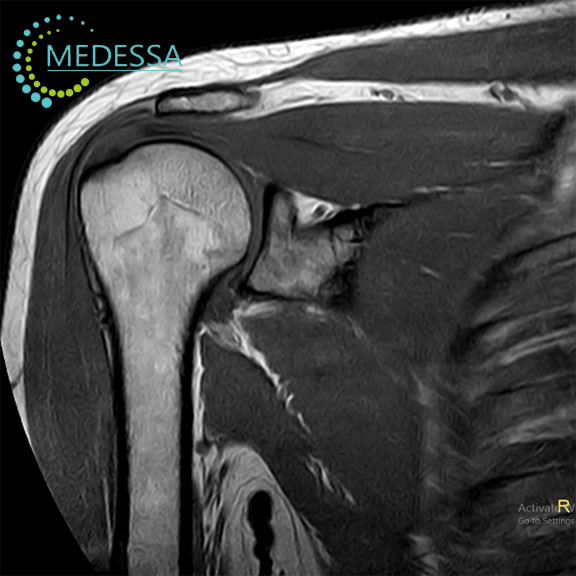
MRI of the Shoulder Joint
Shoulder MRI is a modern diagnostic imaging method that provides detailed visualization of the bones, cartilage, ligaments, tendons, and soft tissues of the shoulder.
The examination uses magnetic fields and radio waves to produce high-resolution images without ionizing radiation. It is widely used in orthopedics, traumatology, and sports medicine.
Indications
Shoulder pain or discomfort
MRI may be recommended for persistent shoulder pain, limited mobility, or unexplained discomfort.
Traumatic injuries
The examination helps detect tendon tears, ligament injuries, fractures, dislocations, and other structural damage.
Inflammatory conditions
MRI can identify arthritis, bursitis, and other inflammatory joint disorders.
Unclear diagnosis
The method is often used when other imaging techniques such as X-ray or ultrasound do not provide sufficient information.
Conditions detected by shoulder MRI
- rotator cuff tendon tears;
- arthritis and degenerative joint disease;
- subacromial impingement;
- shoulder instability;
- soft tissue inflammation;
- benign and malignant tumors.
Common symptoms
- shoulder pain;
- limited range of motion;
- joint swelling;
- clicking or cracking sounds;
- shoulder instability.
MRI of the shoulder joint provides detailed information about joint structures and helps physicians determine the most appropriate treatment strategy.