
MRI of the Knee Joint
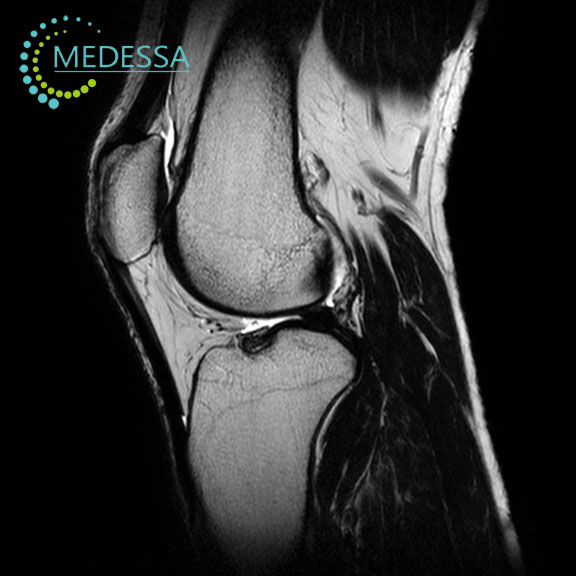
MRI of the knee joint examines the internal structures of the knee using magnetic resonance rather than ionizing radiation. It is particularly useful when symptoms suggest an injury to a meniscus, ligament, cartilage surface or other structure that cannot be adequately characterized by examination and radiographs alone.
The examination is focused on the knee: the distal femur, proximal tibia and fibula, patella and adjacent soft tissues within the prescribed field of view. It is not a complete MRI examination of the thigh or lower leg. The clinical question helps determine which structures need the closest assessment.
What anatomy is assessed on knee MRI?
Menisci and their attachments
The medial and lateral menisci distribute load across the femorotibial joint. MRI depicts their anterior and posterior horns, bodies and root attachments. A meniscal tear may extend to an articular surface; its location, orientation, displaced fragments and associated extrusion can influence interpretation. Intrameniscal signal alone does not necessarily establish a tear, and some subtle lesions remain difficult to identify.
Ligaments and stabilizing structures
The anterior cruciate ligament (ACL) and posterior cruciate ligament (PCL) are assessed for fiber disruption, abnormal orientation and associated injury. The medial collateral ligament (MCL), lateral collateral ligament (LCL), joint capsule and posterolateral corner structures also contribute to stability. MRI findings must be interpreted alongside clinical tests because imaging appearance does not by itself quantify functional instability.
Cartilage, bones and extensor mechanism
MRI depicts the articular surfaces of the femur, tibia and patella, as well as cartilage defects and underlying bone marrow changes. It also evaluates the quadriceps tendon, patellar tendon (patellar ligament), patellar retinacula and infrapatellar (Hoffa) fat pad. The patellofemoral compartment is particularly relevant when symptoms occur during stairs, squatting or knee flexion, although the cause of pain cannot always be determined from MRI alone.
Synovium, bursae and popliteal region
Joint fluid, synovial thickening, periarticular bursae, superficial soft tissues and the popliteal region can be evaluated within the scan coverage. A popliteal (Baker) cyst may accompany intra-articular disease. The appearance of an effusion or synovitis is not specific for a single diagnosis.
When is knee MRI considered?
MRI may be requested after a twisting injury, a suspected meniscal or ligament tear, persistent mechanical symptoms such as catching or locking, recurrent swelling, or unexplained instability. It can also clarify suspected osteochondral damage, an occult or stress-related bone injury, or persistent pain when initial investigations do not explain the symptoms.
In chronic knee pain, radiographs are often the first imaging examination. After acute trauma, the need for radiographs is guided by the injury and clinical findings. MRI is selected when the suspected internal injury or unresolved diagnostic question makes additional soft-tissue or bone marrow information useful; it is not automatically required for every painful knee.
How MRI findings are interpreted
Meniscal injury and mechanical symptoms
MRI may show a horizontal, vertical, radial or complex meniscal tear, a displaced fragment, or a root tear. Meniscal extrusion can accompany root dysfunction or degeneration. Signal changes can also be age-related or postoperative, so the radiologist considers morphology, tear-surface communication, prior surgery and symptoms rather than treating every bright line as a clinically significant rupture.
Cruciate and collateral ligament injuries
Ligament sprains range from edema around preserved fibers to partial or complete disruption. Secondary signs, including characteristic bone contusions or joint fluid, can support the assessment. The posterolateral corner is anatomically complex; subtle injury may require targeted review and correlation with examination.
Cartilage, patellofemoral and marrow abnormalities
MRI can demonstrate focal cartilage defects, osteochondral lesions, marrow edema-like signal and bone contusions. It can help characterize radiographically occult fractures and subchondral insufficiency or stress-related injuries when the clinical findings justify further imaging. In osteoarthritis, MRI can show cartilage loss, meniscal pathology, synovitis and subchondral changes, but it is not routinely needed to establish straightforward radiographic osteoarthritis.
Inflammation, tendons and localized masses
Effusion, synovial thickening, bursitis and abnormalities of the quadriceps or patellar tendon can be seen. Suspected infection, an unusual synovial process or a focal mass may require a tailored examination and further clinical or laboratory investigation. MRI can characterize the extent and tissue relationships of an abnormality but cannot reliably determine every lesion’s cause or histological type.
MRI sequences, imaging planes and contrast phases
A knee examination combines image series acquired in different orientations and with different tissue contrasts. The precise selection depends on the clinical question, equipment, coil and local protocol; the descriptions below are principles, not a mandatory MEDESSA sequence list.
- Sagittal images are especially useful for the cruciate ligaments, meniscal horns, cartilage and extensor mechanism.
- Coronal images help assess the meniscal bodies, collateral ligaments, joint compartments and bone marrow.
- Axial images show the patellofemoral articulation, retinacula, extensor apparatus and selected periarticular structures.
- T1-weighted images provide anatomical detail and help assess normal fatty marrow and marrow replacement patterns.
- PD-weighted and T2-weighted images support assessment of menisci, ligaments, tendons, cartilage and fluid, with diagnostic value dependent on the particular acquisition and orientation.
- PD or T2 with fat suppression (FS), or STIR increase the conspicuity of fluid-sensitive changes such as marrow edema, ligament injury, inflammation and soft-tissue swelling.
A sequence is not a contrast phase. Routine knee MRI is usually performed without intravenous gadolinium. When clinically justified—for example, for suspected infection, a mass, selected inflammatory conditions or particular postoperative questions—additional pre- and post-contrast T1-weighted fat-suppressed images may be used. Dynamic contrast acquisitions are not a standard requirement for routine knee MRI.
Direct MR arthrography is a distinct specialist examination in which contrast is introduced into the joint; it should not be confused with routine MRI or intravenous contrast enhancement. Neither direct arthrography nor optional 3D or diffusion-weighted acquisitions should be assumed to be included in a standard examination.
Knee MRI after surgery
Previous meniscal resection or repair can alter normal meniscal shape and signal, complicating the distinction between healing changes and a recurrent tear. Following ACL reconstruction, MRI may assess graft continuity, tunnel region, associated structures and complications, but graft signal alone does not establish functional integrity. Prior operative reports, earlier imaging and the timing of surgery can materially improve interpretation. Metallic fixation may cause artifacts that obscure nearby tissues.
Preparation and what happens during the examination
Before scanning, patients complete an MRI safety assessment and should disclose implants, surgical hardware, electronic devices, previous operations, possible retained metal fragments and pregnancy. An implant may be MR Safe, MR Conditional or MR Unsafe; its specific identification and manufacturer conditions must be checked rather than assuming all metal is either safe or prohibited.
Usually the patient lies on the scanner table with the knee positioned in a suitable MRI coil or support. Remaining still is important because motion degrades fine anatomical detail. If pain prevents comfortable positioning, inform the radiographer before scanning. The exact duration and image series vary; a fixed examination time cannot be guaranteed.
Intravenous contrast is not usually required for meniscal or ligament assessment. If gadolinium is specifically indicated, relevant prior reactions, pregnancy and kidney-related risk factors are reviewed according to the applicable safety guidance.
What MRI cannot answer on its own
Small or complex tears, very thin cartilage defects, subtle cortical fractures and postoperative changes can be difficult to classify. Motion, metal susceptibility and incomplete field-of-view coverage may reduce diagnostic confidence. MRI abnormalities can be incidental, while symptoms may arise from functional or clinical problems not fully visible on imaging.
Radiographs are valuable for alignment, joint-space narrowing and many fractures; CT can better define selected cortical or complex bony injuries; ultrasound can evaluate superficial tendons and guide some procedures. Arthroscopy allows direct intra-articular assessment when clinically indicated but is invasive and not simply an interchangeable imaging test. The appropriate combination depends on examination findings and the referring clinician’s question.
Related MRI examinations
- MRI of the Lower Leg — for symptoms or suspected injury centered below the knee rather than within the joint.
- MRI of the Thigh — for pathology centered in the thigh outside the standard knee examination.
- MRI of Joints and Soft Tissues — examination catalogue.
Frequently asked questions
Does knee MRI always identify the cause of pain?
No. MRI can reveal structural abnormalities, but some findings are incidental and some pain mechanisms are not visible. The result should be considered together with symptoms and examination.
Can MRI show a meniscal root tear?
It can demonstrate many root tears and associated meniscal extrusion, but subtle lesions may be missed or remain uncertain. The report should be interpreted in the clinical context.
Will a normal MRI rule out knee instability?
Not necessarily. Instability is a functional clinical finding; a normal or equivocal scan does not replace ligament testing and assessment of movement.
Is contrast needed to look for an ACL or meniscal tear?
Usually not. Standard non-contrast knee MRI is generally used for these injuries. Contrast is reserved for specific additional clinical questions.
Can knee MRI be performed after ACL reconstruction?
Often yes, once implant safety is established. Surgical materials may create artifacts, and the operative history helps distinguish expected changes from complications.
Does knee MRI include the whole thigh and lower leg?
No. The field of view is designed around the knee and adjacent structures. A separate examination may be needed for symptoms centered farther along the limb.
Why might I need an X-ray before MRI?
Radiographs can reveal fractures, alignment changes and osteoarthritis. They are often the appropriate first examination for acute injury or chronic knee pain.
What if I cannot fully straighten my knee?
Tell the radiographer before the scan. Positioning may need adjustment, and a restricted position can limit image quality or the evaluation of some structures.
Can MRI distinguish a recurrent meniscal tear after surgery?
Sometimes, but postoperative signal and shape changes can make the answer uncertain. Previous imaging and surgical details are particularly helpful.
Should I bring earlier scans and operative records?
Yes, if available. Prior images, reports and surgical records can help the radiologist interpret chronic, healing or postoperative changes.
Material prepared and edited by: Andrii Bagirov
International medical sources and recommendations
- American College of Radiology — ACR Appropriateness Criteria: Acute Trauma to the Knee.
- American College of Radiology — ACR Appropriateness Criteria: Chronic Knee Pain.
- RadiologyInfo (ACR/RSNA) — Knee MRI.
Last updated
21 October 2026