
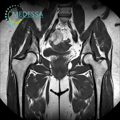
MRI of the Hip Joints
MRI of the hip joints is a modern non-invasive imaging technique used to obtain highly detailed images of the bones, cartilage, ligaments, tendons, and soft tissues of the hip region.
This examination is widely used in orthopedics and traumatology to diagnose inflammatory conditions, degenerative joint diseases, and traumatic injuries.
Indications
Hip pain or discomfort
MRI may be recommended when a patient experiences persistent pain or limited mobility in the hip joint.
Traumatic injuries
The examination can detect fractures, ligament injuries, tendon tears, and other structural damage.
Arthritis
MRI helps evaluate inflammatory joint diseases and the condition of surrounding tissues.
Degenerative disorders
The method can detect osteoarthritis and cartilage degeneration.
Unclear diagnosis
MRI is often used when other imaging methods such as X-ray or ultrasound provide insufficient information.
Conditions detected by hip MRI
- arthritis and inflammatory joint diseases;
- ligament and tendon injuries;
- hip dysplasia;
- avascular necrosis of the femoral head;
- benign and malignant tumors;
- soft tissue abnormalities.
Symptoms that may require MRI
- hip joint pain;
- limited mobility or stiffness;
- clicking or cracking sounds in the joint;
- joint instability;
- weakness in the leg.
MRI of the hip joints provides detailed diagnostic information that helps physicians determine the most appropriate treatment strategy.