
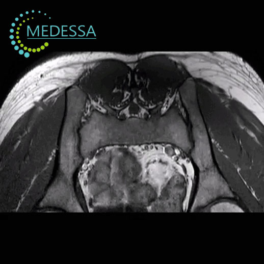
MRI of the Sacroiliac Joints
MRI of the sacroiliac joints is a modern diagnostic imaging method that provides detailed visualization of the sacroiliac joints, pelvic bones, and surrounding soft tissues.
The examination uses magnetic fields and radio waves to produce high-resolution images without ionizing radiation. MRI is particularly useful for early detection of inflammatory joint diseases.
When MRI of the sacroiliac joints is recommended
- suspected sacroiliitis or ankylosing spondylitis;
- genetic predisposition to ankylosing spondylitis (HLA-B27);
- persistent lower back pain;
- inflammatory joint diseases of the lower limbs;
- chronic back pain and reduced mobility;
- reduced flexibility of the spine;
- injuries of the lower back or pelvic bones.
MRI may also be used to monitor the progression of ankylosing spondylitis and other rheumatologic conditions.
Conditions detected by MRI
- intervertebral disc herniation;
- disc protrusion;
- degenerative spine disease;
- spinal canal stenosis;
- inflammatory processes;
- spinal tumors;
- post-traumatic changes.
Indications for MRI
- neck pain;
- headache or dizziness;
- numbness or weakness in the arms;
- limited neck mobility;
- suspected disc herniation;
- spinal injuries;
- suspected tumors or inflammatory diseases.
MRI of the cervical spine provides detailed visualization of spinal structures and helps physicians determine an accurate diagnosis and treatment plan.