
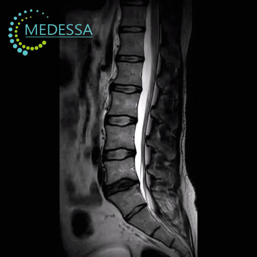
MRI of the Lumbar Spine
MRI of the lumbar spine is an advanced diagnostic imaging method used to evaluate the lumbar vertebrae, intervertebral discs, nerve roots, and surrounding soft tissues.
The examination uses magnetic fields and radio waves to produce detailed images without ionizing radiation. MRI is one of the most informative methods for diagnosing spinal disorders.
Structures evaluated during MRI
Vertebrae
The condition, structure, and alignment of the lumbar vertebrae are assessed to detect fractures, deformities, or other abnormalities.
Intervertebral discs
MRI can identify disc herniation, disc protrusion, and degenerative disc disease.
Ligaments and muscles
Soft tissues, ligaments, and muscles surrounding the lumbar spine are evaluated.
Spinal canal
The examination helps detect narrowing or compression of the nerve roots.
Conditions detected by MRI
- intervertebral disc herniation;
- disc protrusion;
- degenerative spine disease;
- spinal osteoarthritis;
- inflammatory disorders;
- spinal tumors;
- post-traumatic changes.
Indications for MRI
- lower back pain;
- radiating leg pain;
- numbness or weakness in the legs;
- limited spine mobility;
- suspected disc herniation;
- spinal injuries;
- suspected tumors or inflammation.
MRI of the lumbar spine provides detailed visualization of spinal structures and helps physicians determine an accurate diagnosis and treatment plan.