
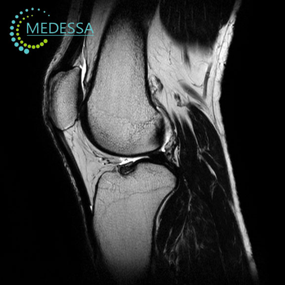
Knee MRI
MRI of the knee joint is a modern non-invasive imaging technique that provides highly detailed visualization of the internal structures of the knee.
The examination uses magnetic fields and radio waves to produce images without ionizing radiation. MRI is widely used in orthopedics and sports medicine to diagnose injuries and diseases affecting the knee joint.
Structures evaluated during knee MRI
Articular cartilage
Articular cartilage covers the ends of bones and allows smooth joint movement. MRI can detect cartilage damage, thinning, and degenerative changes.
Menisci
The menisci are crescent-shaped cartilage structures located between the femur and tibia. They act as shock absorbers and help stabilize the joint. MRI is highly effective in detecting meniscal tears and degeneration.
Cruciate ligaments
The anterior and posterior cruciate ligaments stabilize the knee joint. MRI allows accurate evaluation of ligament injuries, including partial or complete tears.
Indications
Knee pain or instability
MRI is used to determine the cause of persistent knee pain or limited mobility.
Traumatic injuries
The examination helps diagnose meniscal tears, ligament injuries, and other internal joint damage.
Inflammatory conditions
MRI can detect arthritis, synovitis, and other inflammatory diseases of the knee.
Degenerative disorders
The method helps diagnose osteoarthritis and cartilage degeneration.
Advantages of MRI
- high-resolution imaging;
- detailed assessment of soft tissues;
- multiplanar visualization;
- no ionizing radiation;
- non-invasive examination.
MRI of the knee joint is one of the most accurate methods for diagnosing knee injuries and joint diseases.