
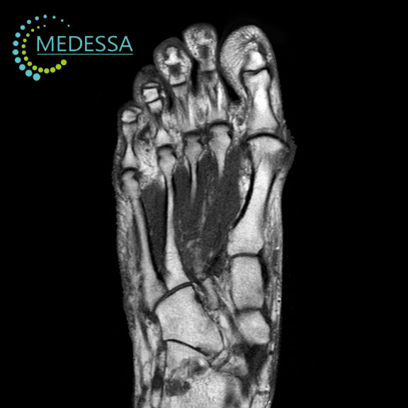
MRI of the Distal Foot
MRI of the distal foot is a non-invasive imaging technique used to obtain detailed visualization of the bones, joints, ligaments, tendons, and soft tissues of the forefoot.
The examination is widely used in orthopedics and sports medicine to diagnose injuries, inflammatory conditions, and degenerative disorders.
Indications
Foot pain
MRI helps determine the underlying cause of persistent foot pain.
Traumatic injuries
The method can detect fractures, ligament injuries, tendon tears, and other soft tissue damage.
Joint disorders
MRI is used to diagnose arthritis, gout, osteoarthritis, and plantar fasciitis.
Stress fractures
The examination may reveal microfractures and inflammatory changes in bone or soft tissue.
Advantages of MRI
- high-resolution imaging;
- excellent visualization of soft tissues;
- multiplanar imaging capability;
- no ionizing radiation.
MRI of the foot is a valuable diagnostic tool that helps physicians accurately assess foot injuries and diseases.