
MRI of the Male Pelvic Organs
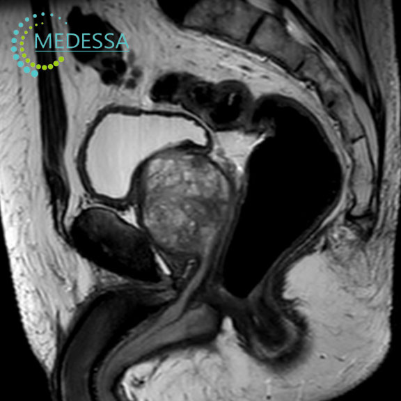
MRI of the male pelvic organs is a non-invasive imaging technique used to obtain detailed visualization of the prostate gland, seminal vesicles, urinary bladder, lymph nodes, and surrounding tissues.
The examination is widely used in urology to detect inflammatory conditions, tumors, and other pelvic abnormalities.
Indications
Pelvic pain or discomfort
MRI helps determine the cause of pain or discomfort in the pelvic region.
Structural abnormalities
The method allows evaluation of the prostate, seminal vesicles, and urinary bladder.
Suspected tumors
MRI is commonly used to detect prostate and bladder tumors.
Post-treatment follow-up
The examination can monitor treatment outcomes and disease progression.
Conditions detected by MRI
- prostatitis;
- benign prostatic hyperplasia;
- prostate cancer;
- pelvic trauma;
- bladder tumors.
MRI of the male pelvis is an important diagnostic tool that helps physicians establish an accurate diagnosis and plan appropriate treatment.